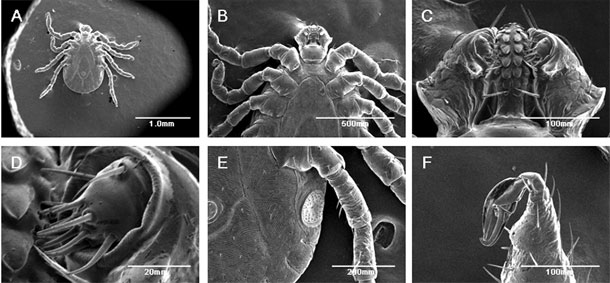
You didn’t wake up this morning thinking that a tick under a scanning electron microscope was going to be the coolest thing you saw all day, and yet here you are. After discovering some ticks alive inside a vacuum drying chamber, Yasuhito Ishigaki of Kanazawa Medical University decided to see if the hardy little bloodsuckers could stand up to the electron bombardment and vacuum conditions inside a scanning electron microscope (SEM). They could, and he’s got the video to prove it.
SEM rigs are great for capturing very fine detail of very small things, but they aren’t easy on their subjects. They work by bombarding a sample with electrons and recording how they scatter to create an image. Air interferes with this electron beam, so all this takes place inside a vacuum. And samples are often stained or even coated with metal beforehand to enhance the resolution of the microscopy.
All said, life is not good for a SEM sample. In fact, putting anything living into an SEM sample chamber pretty much ensures that it won’t be living when you take it out. But this clearly isn’t true for ticks. In the video below, you can clearly see the tick moving its legs. Ishigaki did this with 20 different ticks, and all of them survived, making them the first animals to ever be scanned with SEM.
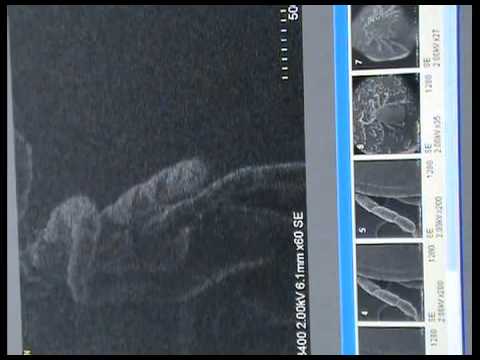
Living tick filmed under an electron microscope
