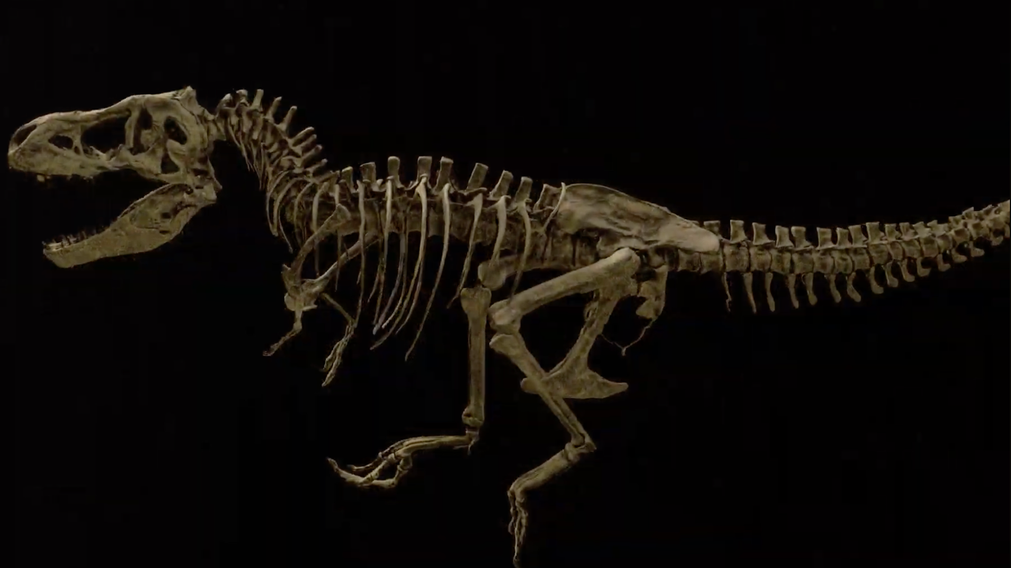
The largest Tyrannosaurus rex fossils ever discovered belong to a 66-million-year-old apex predator named Scotty. Like many T. rex skeletons, it’s difficult to determine if the specimen was male or female, but Scotty was an unequivocal bruiser. Standing 13-feet-tall at the hip and measuring upwards of 43-feet-long, the Late Cretaceous hunter weighed as much as 19,500 pounds.
However, Scotty isn’t simply the biggest T. rex on record. One of its bones still has traces of preserved soft tissue. This is almost never seen in paleontology, as soft tissues degrade due to the ravages of time. The only way to safely examine these delicate remains is through highly sensitive techniques like neutron imaging. But with the help of researchers at the Department of Energy’s (DOE) Oak Ridge National Laboratory (ORNL) in Tennessee, paleontologists recently glimpsed Scotty’s rib and its remaining tissue in unprecedented detail. The results offer some of the first in-depth looks at fossilized dinosaur tissue on a microscopic level.

ORNL Neutron Bone Imaging Brings T. Rex to Life
“It’s like winning the lottery,” Mauricio Barbi, a physicist at the University of Regina (U of R) in Saskatchewan, Canada, said in a recent interview. “Scotty’s rib contains a vast network of mineralized blood vessels that has never before been observed in a fossil.”
The reason for the blood vessels’ preservation is also a window into the T. rex’s death. The broken rib indicates that Scotty likely succumbed to injuries sustained during a dinosaur brawl. After the rib cracked, iron-saturated blood covered the wound and formed tiny blood vessels to facilitate faster healing. The anatomical response wasn’t enough, and Scotty soon died in a salty marsh. The surrounding environment slowed down the T. rex’s decomposition so much that it allowed many of those blood vessels to fossilize.
To get a closer look at these details, paleontologists relied on neutron imaging to see the lighter elements present in the fossils as well as X-ray imaging to look at the heavier ones. From there, researchers also used synchrotron radiation and other microscopy approaches to analyze the healing tissues on a cellular level. Importantly, all of this imaging was possible without damaging Scotty’s priceless fossils.
“Neutrons not only corroborated what we found with synchrotron radiation techniques that led to the discovery of blood vessels in Scotty’s rib,” explained U of R physicist Marcella Berg. “They also proved to be a highly valuable addition to our current studies in search of soft tissue preservation in fossils.”