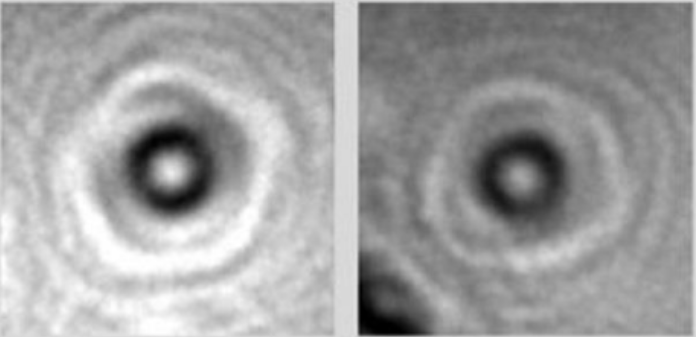

There are thousands of different proteins in the human body. Each has a unique shape that determines its function. Scientists have a hard time capturing images of individual proteins, however—the high-powered imaging tools would obliterate the fragile proteins, so researchers capture photos of proteins in a crystal structure, often millions of them at a time. The resulting images are often blurry, and some proteins can’t be photographed because they don’t form crystals. Now a team has used wonder-material graphene to take the first photos of individual proteins, according to a study published recently on arXiv and reported by New Scientist.
To capture an image of a single protein, the researchers spray a mixture of proteins in solution onto a thin sheet of graphene. They then used a low-energy holography electron microscope, which creates an image by bouncing a beam of electrons off the proteins, then recording how those electrons interact with a pattern of other electrons. That low energy ensured that the protein wasn’t obliterated while the researchers were taking its photo. Using a computer, the researchers used the hologram image to reconstruct the protein’s original structure.

The researchers tried this imaging technique with a few proteins with well-known structures: hemoglobin (the protein that carries oxygen in red blood cells), bovine serum albumin (a cow protein commonly used in lab experiments), and cytochrome c (proteins used to transfer electrons in the body). They compared the resulting images to those taken from other imaging techniques and found that their photos had less blurring. The researchers next hope to take photos of proteins that have never before been seen on their own. If scientists better understand proteins’ structure, they may be able to figure out what goes wrong in diseases linked to misfolded proteins, such as Alzheimer’s, Parkinson’s, and Huntington’s.
