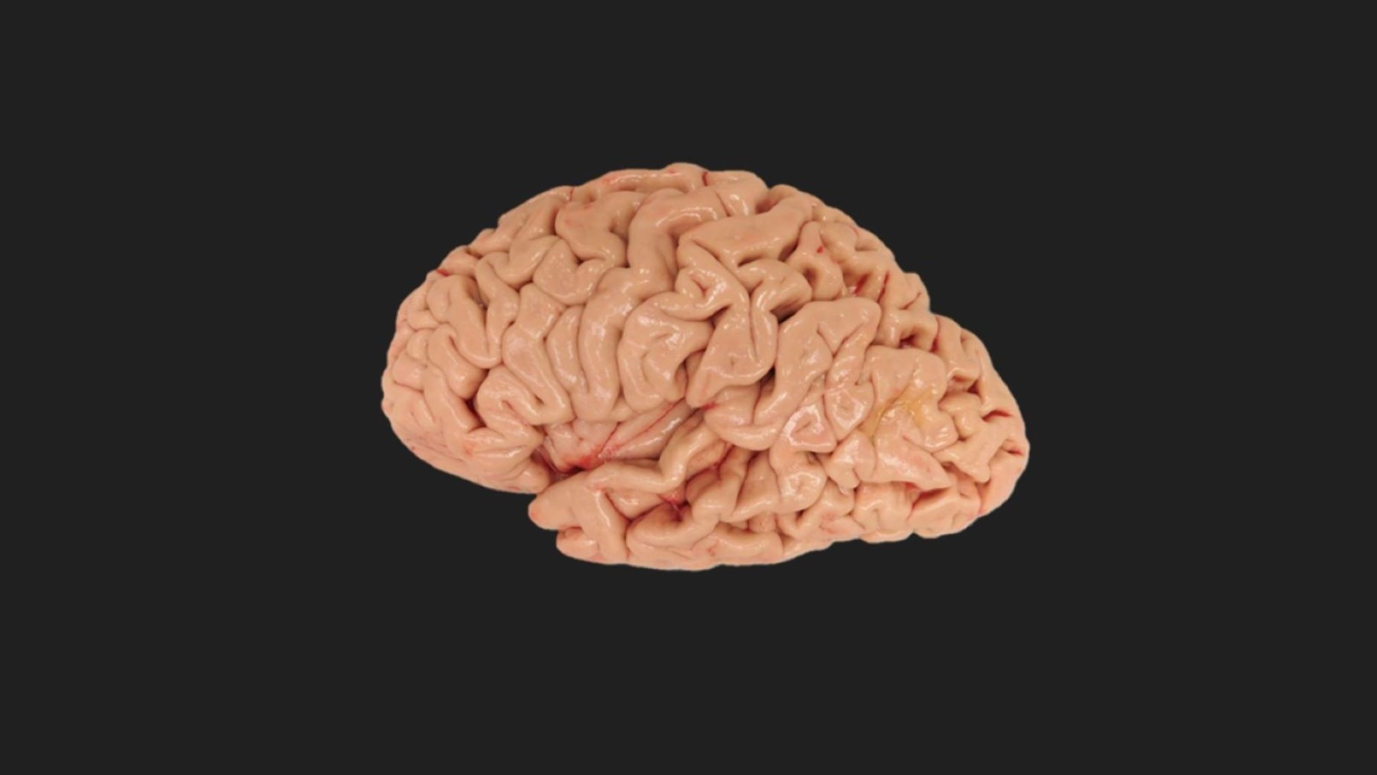

We’re closer than ever to mapping the entire brain to the microscopic level. Hundreds of neuroscientists across the world recently characterized more than 3,000 human brain cell types as part of the National Institute of Health’s BRAIN Initiative Cell Census Network, publishing almost two dozen papers in four Science journals today. This super-focused attention to detail could unlock many mysteries surrounding that complex organ, such as what happened in our brains to distinguish us from other primates.
“This is the first large-scale, detailed description of all the different kinds of cells present in the human brain,” says Rebecca Hodge, an assistant investigator at the Allen Institute in Seattle who co-authored multiple studies in the paper package. Her hope is that this brain atlas provides a community resource for scientists to explore how the wide variety of brain cells contribute to health and disease.
Mark Mapstone, a professor of neurology at University of California, Irvine School of Medicine, who wasn’t involved with these studies, likened the new data about the brain to a tourist’s guide. “Imagine navigating an unfamiliar city with a roughly drawn street map containing only the major streets of the downtown compared to navigating the same city with a detailed map extending beyond the downtown to the suburbs and including all highways, two-way and one-way streets, alleyways, sidewalks, location of street signs and traffic signals, speed limits, and location of coffee shops and restaurants,” he says. “Cleary, the latter would make navigation and understanding the city much easier.” This first suite of studies shows three main ways the brain map can be used for biology and medicine.
An evolving brain
A human brain atlas can teach us about our evolutionary history. One study published today in Science used single-nucleus RNA sequencing to measure the gene expression of individual brain cells in humans and five other primate species, including chimpanzees and gorillas. In this method, scientists pull out individual cells from a piece of tissue, break them open to expose the genetic messengers inside, then use tags akin to tiny barcodes to identify that material. “This is the main technology used in some of these papers that are coming out and it’s a technique that’s only been around for the past 10 years,” Hodge says. Getting this genetic profile allows researchers to group clusters of cells into specific types.
[Related: Psychedelics and anesthetics cause unexpected chemical reactions in the brain]
Our cells’ composition and organization is similar to those of our close relatives. However, the biggest differences seemed to occur in a brain region called the middle temporal gyrus, which is involved in processing semantic memory and language. Humans had higher numbers of projecting neurons in this area compared to other species. What’s more, the researchers highlighted a difference in gene expression that promoted synaptic plasticity, which is the ability of neurons to strengthen brain connections. This feature is an important component for learning and memory, and it might explain how humans developed complex cognitive skills.

There was some variation within humans, too. Another study found the most differences across humans in immune cells called microglia as well as deep-layer excitatory neurons, which are involved in the communication between distant brain regions. Researchers are not quite sure why—one theory is that deep-layer excitatory neurons develop earlier and are more exposed to environmental factors that could diversify their gene patterns. “Everyone’s brain is largely similar. Even though we have the same building blocks, it’s the small number of differences that matter,” says Jeremy Miller, a senior scientist at the Allen Institute, and co-author of the study. “We’re now starting to understand how important these changes are and figuring out what makes us uniquely human.”
Animal models
Because human brains share many features with other mammals, neurologists frequently use the small brains of mice to study diseases. The one problem, Miller says, is that mice don’t naturally develop neurodegenerative diseases common in humans. Scientists who want to study Alzheimer’s disease, for example, would need to manipulate multiple mouse genes to cause the kind of brain pathology seen in older people. This requires a comprehensive understanding of how cell types in the brain work together and how they change in the context of disease.
[Related: How your brain conjures dreams]
Much brain research in mice focuses on the neocortex, responsible for higher cognitive function. It might seem reasonable to assume that much of the brain’s cellular complexity appears here. But this doesn’t seem to be the case. In one of the first studies to create a cell map of the entire adult brain, neuroscientists have found high levels of diversity in older evolutionary structures such as the midbrain, which is involved in movement, vision, and hearing, and the hindbrain, which governs vital bodily functions such as breathing and heart rate. In subcortical areas, there also appears to be a supercluster of cells called splatter neurons that control innate behaviors and physiological functions. Replicating the complexity of these particular brain regions in animal models could help better identify the cellular origins of human diseases.
Personalized medicine
Imagine a future where treatments are tailored to someone’s specific needs. To do that, scientists would use a person’s genetic profile, rather than characteristics such as weight or age, to inform any medical decisions. Clinicians could also use this genetic information to identify the risks of potential diseases and provide early preventative measures.
“A detailed brain atlas can help us understand what successful brain function looks like so we can maximize brain cells and circuits that promote brain heath,” Mapstone says. “Addressing brain disease and promoting brain health can be more easily accomplished if we know how these cells are organized. “

Doctors are already using people’s genetic information to assess whether patients would be good candidates for a particular cancer treatment or to find the proper dose of a drug. This may soon include testing for neurological conditions. One study, which analyzed 1.1 million cells in 42 brain regions of neurotypical adults, identified specific neuronal cell types—mainly in the basal ganglia, a region involved in addictive behaviors—that were linked to 19 neuropsychiatric disorders and traits. Those conditions included schizophrenia and bipolar disorder as well as alcohol and tobacco use disorder.
This project is a step in the right direction for advancing research in personalized medicine, says Miller, though he warns this is only one of many to make this a reality for everyone.
Miller and Hodge are optimistic there will be other versions of the human brain atlas completed in the next five years, as other groups wrap up similar projects.
But there’s a possibility that we’ll never get the full picture. While Miller finds a half-decade timeframe reasonable, he says there’s always a chance science develops a new technology that could unearth something unexpected about the brain. “We can always do more,” he says.
This post has been updated.
