The openVertebrate project was a five-year initiative funded by the National Science Foundation to make 3D models of museum specimens freely available to scientists, students, teachers and the public.
openVertebrate
Get the Popular Science daily newsletter💡
Breakthroughs, discoveries, and DIY tips sent six days a week.
By signing up, you confirm you are 16+, will receive newsletters and promotional content and agree to our Terms of Use and acknowledge the data practices in our Privacy Policy. You may unsubscribe at any time.
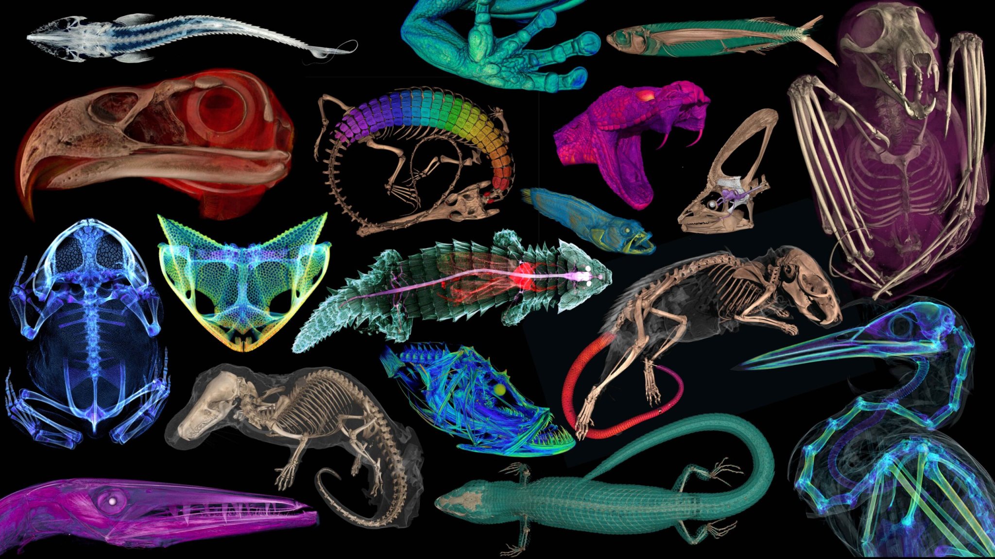
If dissecting a frog in biology class had you begging to be sent home, new 3D scans of thousands of vertebrate species are here to help by letting you peek at animal insides without the mess. The newly completed openVertebrate (oVert) project took five years and brought together 18 natural history institutions to create the free online museum, showing the anatomy and physiology of over 13,000 specimens. A summary of the work was published March 6 in the journal BioScience.
From 2017 to 2023, oVert project members took detailed CT scans of more than half the genera of all amphibians, reptiles, fishes, and mammals. The scanners used high-energy X-rays to look past the organism’s scales, fur, or skin to view the dense bone structure beneath. Scientists stained some of the specimens with a temporary contrast-enhancing solution that allows the team to visualize their soft tissues, including muscle, skin, and other organs.
“Museums are constantly engaged in a balancing act,” David Blackburn, principal investigator of the oVert project and curator of herpetology at the Florida Museum, said in a statement. “You want to protect specimens, but you also want to have people use them. oVert is a way of reducing the wear and tear on samples while also increasing access, and it’s the next logical step in the mission of museum collections.”
Take a look at some of the incredible scans below. It will be like stepping back into high school biology, without the scalpel, Bunsen burners, or safety glasses.
An analysis of oVert specimens revealed that frogs have lost their teeth over 20 times throughout their evolutionary history, more than any other vertebrate group. Image: openVertebrateWith CT scanning, scientists can study a specimen’s internal anatomy without the need for dissection. Image: openVertebrateThe primary goal behind the oVert project was to image as great a breadth of diversity across the vertebrate tree of life as possible, including fish, reptiles, amphibians, birds and mammals. Image: openVertebrate Osteoderms have evolved multiple times in different animal groups, and their presence in spiny mice further indicates the genetic pathways needed to produce them are highly conserved among vertebrates. Image: openVertebrate Researchers performed a digital dissection of North America’s rarest snake when a specimen was found that had died while trying to eat a centipede. Image: openVertebrate The preserved specimen of a black bellied fruit bat compared to its CT scan. Image: openVertebrate.The gopher tortoise’s internal anatomy. The colorful regions show soft tissues, including organs. Image: openVertebrate.Caralophia loxochila, or the Slantlip eel swims in the shallow waters of the western Atlantic Ocean, near Brazil, the Bahamas, and the Florida Keys. Image: openVertebrate.A model of a Komodo dragon’s skull. Bites from their powerful jaws can be fatal to humans. Image: openVertebrate.Multiple snake species that were temporarily stained with an iodine solution showing their soft tissues before being scanned. Image: openVertebrate.