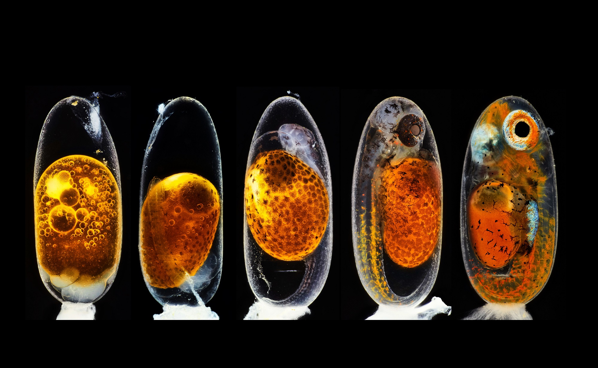

Microscopes were invented a good 300 years before photography came into fashion. But it still took a few decades for someone (more specifically, a surgeon in the US Army) to combine the two and make science shine brighter in the public eye.
A century and a half later, researchers are still honing their artistic skills through photomicrography. From cancer cells to newly budding life, the perspectives captured by pairing powerful lenses offer a level of beauty and technical detail that are hard to come by. The Nikon Small World awards make it so that the sharpest, most unique visuals get some spotlight outside of the lab. PopSci picked out a few of this year’s still image winners for you to take a guess and gander at.
Seen through a 10x objective microscope lens, this subject experiences quick growth over a nine-day period. That should make it clear that it’s not a fish-oil pill (though close!).
Click to see the Answer
A clownfish embryo
With confocal imagine, we get 3D-reconstructed views of this grainy specimen’s cross sections. The colors are a computerized touch as well.
Click to see the Answer
Pollen from a hebe shrub
Heating up an ethanol and water solution led to this otherworldy formation. The image was captured through polarized-light filters to enhance the structure.
Click to see the Answer
Amino acids glutamine and beta-alanine
Zoomed in at 40x, this toothy body part plays a crucial role in one slimy animal’s daily life. Our human version looks much less metal.
Click to see the Answer
Snail tongue
It’s obvious that this is a skeleton of sorts. But the simple brightfield approach, which plays on illumination and reflection, gives the complex bone structure an air of mystery.
Click to see the Answer
Fruit bat skeleton
Sometimes the most unassuming subjects prove to be the best muses. At 9x magnification, this everyday accessory seems more rugged and complex than you’d expect.
