
The discovery of DNA as a double helix is a hallowed story of scientific triumph—the work of four researchers merging together to solve one of science’s biggest mysteries, giving birth to what we know as the field of modern genetics. But decades later, we’re still learning that DNA is a more furiously complicated piece of biological machinery than we ever knew.
For the first time ever, scientists have just discovered a new shape of DNA lurking inside human cells. In a study published Monday in Nature Chemistry, a team of researchers from the Kinghorn Centre for Clinical Genomics at the Garvan Institute of Medical Research in Sydney describe finding DNA as a four-stranded knot-like structure—called an i-motif—in human cells, upending much of what we previously thought could and could not exist in living humans, and eliciting a slew of questions about what the role of this structure might be, if it even has one.
We already know DNA can come in other forms, such as triple helices or cruciforms. And the i-motif is not the first four-stranded structure to be found in human cells; scientists already did that with the discovery of G-quadruplex DNA in humans in 2013. But this is the first time i-motifs have ever been found in human cells. The i-motif structure was first observed around two decades ago, in lab conditions that were quite acidic, and most assumed the i-motif would probably never be found in nature.
“This raised a scientific debate concerning the biological relevance of this motif,” says Daniel Christ, director for the Centre for Targeted Therapy at the Garvan Institute and a co-author of the new study. “We are providing first direct evidence that the i-motif structure exist in cells under physiological conditions.”
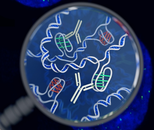
Here’s how the i-motif works: imagine a small section of the DNA double helix where the hydrogen bonds that connect the two major strands come apart while the helix suddenly untwists. If one of the strands is chock-full of cytosine (one of the four major nucleic acids that comprise DNA), it will loop outward like a tied shoelace. Hydrogen bonds form within the loop itself, binding those cytosines to one-another (instead of to guanine, as is normally the case in the double helix).
“They essentially form a scaffold, where each C-C bond is 90 degrees to its corresponding C-C pair,” says Laurence Hurley, a medical chemist at the University of Arizona who has also studied i-motifs.

To confirm the presence of these i-motifs in human DNA and pinpoint their locations, the Sydney team created a special fragment of an antibody molecule capable of binding to the i-motif structure. They then used fluorescent techniques to highlight the antibody molecules under the microscope. It’s a very tried-and-true method in chemistry and biology, and should wash away lingering doubts about the veracity these i-motifs can arise in nature.
But what does the i-motif do? There’s quite a bit of evidence to suggest it plays a role in transcription (when the cell uses DNA as instructions to make different proteins). The Sydney team studied the presence of i-motifs during all phases of the cell cycle, and found that the motifs appear most commonly when DNA is being actively transcribed, but disappear when DNA is being replicated.
They also found that the motifs often appeared in parts of the promoter regions of genes, which are not read and expressed as protein products, but instead can turn on and off the expression of other genes and prevent or promote the production of certain proteins. “We consider it likely that the formation of i-motifs in promoter regions fine-tunes the expression of corresponding genes,” says Christ.
According to Hurley, there are specific proteins and mechanisms that work to unwind the DNA, create the i-motif folds and stabilize them during transcription, and then unfold these knotty loops and zip things back up into a double helix when it’s time for cell division. And this can be accomplished without the super-acidic setting typically needed for these motifs. “That’s where the power of these structures are,” he says. “They are highly dynamic, and you can fold them and unfold them, in order to activate transcription.”
“It’s clear that these structures [i-motifs] are implicated in gene expression,” says Hurley. “This paper provides the icing on the cake.”
Hurley and others have previously found evidence that i-motifs are related to a number of cancer-related genes, like MYC (expressed in over 80 percent of cancers), KRAS (which controls cell growth and proliferation signaling), and BCL-2 (which prevents cancer from undergoing apoptosis, or programmed death). Hurley himself recently founded a new company, Reglagene, that is seeking to use these i-motifs as potential targets for new cancer drugs, and prevent oncogenesis at the genetic level itself, as opposed to “undruggable” protein targets.
While the new findings pretty solidly show i-motifs can show up in living human cells, Hanbin Mao, a biochemist at Kent State University who was not involved with this study, notes there is room for more convincing evidence that i-motifs are a natural phenomena. The Sydney researchers cannot say for sure the antibody wasn’t binding to other targets, and more importantly, that the antibody’s binding to the DNA did not promote the formation of i-motif itself. Needless to say, there are years or even decades’ worth of follow-up research in store to learn more about what i-motifs are, how they work, why they exist, and how we might be able to harness their powers.
Nearly 65 years after Watson, Crick, Franklin, and Wilkins, the DNA plot continues to thicken.
