Not every mummy is treated equally. While the traditional image conjures a well-preserved, carefully wrapped ancient Egyptian body inside an elaborately decorated tomb, there are many more examples of partial and poorly prepared remains. These especially delicate specimens are difficult for scientists to even document, much less analyze in sufficient detail.
Take a collection of mummy fragments housed at the MNMKK Semmelweis Museum of Medical History in Budapest, Hungary. Curators have housed the archaeological discoveries since the institution opened in 1965, and at least some of the mummies are over 2,300 years old. But apart from the radiocarbon dating, they lacked the technological capabilities to safely study them. However, researchers can finally see the remains with impressive clarity thanks to the recent installation of a high-resolution CT scanner.

“Based on the results so far, it is evident that modern imaging technology opens up new perspectives in mummy research. It can reveal information hidden in finds that are thousands of years old without damaging them,” collection curator Krisztina Scheffer said in a statement.
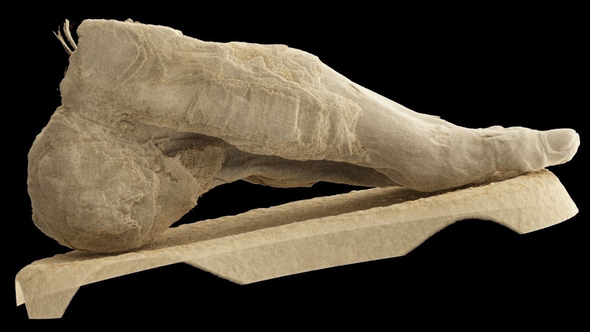
The results are a great reminder that even experts don’t always get it right the first time. Want proof? One specimen previously believed to be a mummified human head—or possibly even a bird—turned out to be an adult’s foot.
The post-mortem imaging team also looked at teeth, two heads’ skull sutures, and an individual’s partial limbs. In the latter case, they now think the person was relatively young and suffered from osteoporosis, but they remain unsure about the cause. That said, the museum team is confident that these findings are only the beginning.
“The current images provide a more detailed view than ever before,” said Scheffer, adding that further work is, “expected to yield new, scientifically valid findings regarding the remains that have been preserved in the collection for decades.”
Take a look at some of the other images below.


