A new lab technique now lets scientists make gorgeous, fully intact images of bodily organs such as the brain.
A team of engineers has developed a way to turn organs from mammals, such as lab mice or human bodies donated to science, transparent. Once transparent, scientists can add chemicals to the organs that attach to and highlight specific features, such as different cell types. The result is an intact organ that scientists can see inside and study.
Because such visualization can be great for whole-organ studies, this isn’t the first time scientists have tried to make some transparent brains. This new technique, called CLARITY, works better with chemical labels and is quicker than previous techniques, CLARITY’s developers wrote in a paper they published today in the journal Nature.
To demonstrate their technique, the engineers, who are all from Stanford University, imaged some mouse brains:


Or check out this Nature video to see some more images, plus some rotations:

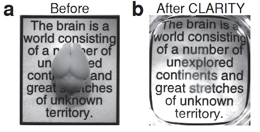
See-through brains
Making these images is an eight-day process. The Stanford researchers started by infusing a mouse brain with a hydrogel solution. They then put the gel and brain into an incubator to set. (Like making Jell-O! Except that the setting, in this case, required a higher temperature rather than a lower one.)
The set gel bound to and physically supported most of the things in the brain. The gel didn’t bind to lipids, or fats, in the brain, however. Such fat is opaque and surrounds each cell. When researchers extracted this unbound fat, they were left with a clear view of everything else, frozen in place. For example, proteins that were originally embedded in cell membranes and the little spines that come off of neurons both remained.
At this point, the researchers could add different molecules to color the parts of the brain they want to study and look at the whole thing under a light microscope.
