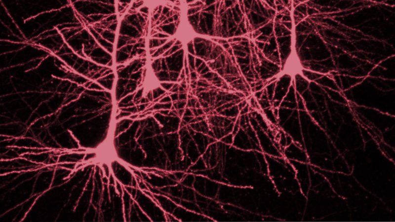
We love it when science and art intersect. Sometimes that happens in the form of a stunning infographic, a colorful map, or a well-timed photograph. Microscopic imaging in particular provides some of the most remarkable insights into worlds strange and unseeable to the naked eye.
Every year, the Olympus BioScapes competition celebrates achievements in light microscopy and the scientific insights they provide. Scientists from around the world submit their photos and videos, and a team of PhD-toting judges pick their favorites. The entrants are judged “based on the science they depict, their beauty or impact, and the technical expertise involved in capturing them.” And this year’s winners [have just been announced](https://www.olympusbioscapes.com/content/pressreleases/BioScapes/ 2014 Press Release.pdf)!
First prize goes to a group of researchers who used a novel imaging technique to track every cell in a developing fly embryo. The technique could help scientists better understand how animals develop, which could in turn aid in understanding diseases. At the end of the video, you can see the freshly built larvae crawl off-screen:
Researchers from the HHMI Janelia Research campus in Virginia used a custom setup to capture neural activity in a living zebrafish. The video shows 3-D recordings of the brain signals from approximately 100,000 neurons.
We look again at the zebrafish brain, this time using an imaging technique that picks up on the calcium that each neuron releases when it’s activated during the perception of juicy prey.
Here’s a beautiful video of Volvox rousseletii, a type of algae, with embryos developing inside it. Each embryo turns itself inside out at the end of embryogenesis. A team from Tokyo’s Chuo University captured the process using confocal and differential interference contrast microscopy.
Watch the 3-D reconstruction of a zebrafish’s beating heart in this video from the Max Planck Institute:
This next one is great because … bears. Water bears, that is. Called tardigrades, these little guys can survive practically anything, including, we think, the extreme conditions of outer space. Here you can see one lumbering around upside-down.
For more amazing microscopy imagery, be sure to check out the gallery of winners and honorable mentions. You can also scroll through our gallery of the winners from the Nikon Small World microscopy challenge.