Ultra-Powerful New Microscope Displays Molecules and Cell Movement In Real Time
For scientists studying the smallest components of life, microscopes have always had frustrating limitations. Electron scanning microscopes can see very...
For scientists studying the smallest components of life, microscopes have always had frustrating limitations. Electron scanning microscopes can see very small object, but not in real time through the dynamic movement of cells. Fluorescent dyes identify microscopic objects, but the brightness of the emitted light greatly reduces the resolution.
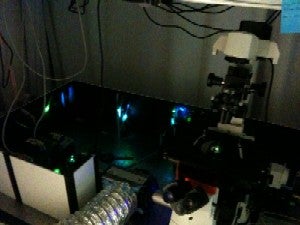
The Stochastic Optical Reconstruction Microscope (STORM) solves both those problems. 100 times more powerful than a regular optical microscope, the STORM filters and adjusts light emitted from fluorescent dyes to produce a clean image of individual molecules, and thus allowing researchers to watch the behavior of proteins in real time.
Stochastic Optical Reconstruction Microscope
Basically, STORM lets researchers make a video of any of cell’s behavior, be it DNA replication or ATM synthesis, in definition so high they can see the individual components of each cellular organelle. While an older light microscope could only identify the center of a protein to within 0.2 inches, the STORM can identify that center to within two billionth of an inch.
Developed by Jennifer Ross, a physics professor at the University of Massachusetts, Amherst, the STORM relies on a new fluorescent tagging technique that gives researchers the ability to modulate the light emitted from a tagged molecule. Progressively activated and deactivated adjacent fluorescent proteins provide a contrast that allows the STORM creates composite, mosaic, images with a greater resolution than traditional dye images, and in living, moving cells in real time.
Unfortunately for those biologists though, they’re going to have to wait a little while before getting to play with this new toy. The STORM still has two more years of testing with the physics department before those biologists get a crack at it.