SHARE

Nikon’s annual Small World Competition began in 1974 to showcase the best microscope-aided photography. The competition attracts a fascinating variety of subjects, photographed using a range of microscopy techniques. Many of the images are scientifically important, but all are aesthetically stunning. This year’s 137 winners have been announced, and we’ve got them all here for you.

View photo gallery

This strangely glowing, somewhat translucent shot captures the anther of a thale cress, an organism whose genome was fully sequenced in 2000. According to photographer and research scientist Dr. Heiti Paves, the photo has no scientific significance—it just happened to be the most artistic in his pile of thousands. Still, the thale cress is known to be a model organism in plant biology.

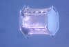
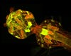
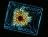
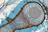
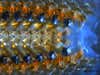
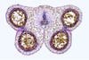
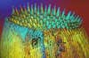
1st Place: Arabidopsis thaliana (thale cress) anther (20x)

This strangely glowing, somewhat translucent shot captures the anther of a thale cress, an organism whose genome was fully sequenced in 2000. According to photographer and research scientist Dr. Heiti Paves, the photo has no scientific significance—it just happened to be the most artistic in his pile of thousands. Still, the thale cress is known to be a model organism in plant biology.
Who knew a plant with small yellow flowers, spiny leaves and the potential to reach six feet in height could make for such a pretty picture? Cut the stem of this invasive weed and a milky sap will flow out, but you won't find that in the part pictured here. Although the dandelion-like blossoms are yellow, the stem boasts a range of colors hidden to the naked eye.

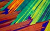
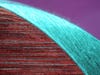
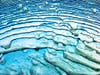
2nd place: Sonchus asper (spiny sowthistle) flower stem section (150x)

Who knew a plant with small yellow flowers, spiny leaves and the potential to reach six feet in height could make for such a pretty picture? Cut the stem of this invasive weed and a milky sap will flow out, but you won’t find that in the part pictured here. Although the dandelion-like blossoms are yellow, the stem boasts a range of colors hidden to the naked eye.
When objects like circuits are going to be chemically etched, they are often first covered with photoresist. Photographer Dr. Pedro-Barrios Perez inspects the photomechanical substance to make sure it will work when it goes to production. So why did he submit this picture? "It looks like the sun warming up the earth with its mighty energy waves," he says. Interesting, because photoresist sometimes wrinkles when exposed to heat.

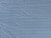
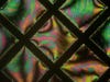
3rd place: Wrinkled photoresist (200x)

When objects like circuits are going to be chemically etched, they are often first covered with photoresist. Photographer Dr. Pedro-Barrios Perez inspects the photomechanical substance to make sure it will work when it goes to production. So why did he submit this picture? “It looks like the sun warming up the earth with its mighty energy waves,” he says. Interesting, because photoresist sometimes wrinkles when exposed to heat.
This cross-section of an anglerfish's ovary is reminiscent of another sea creature—the nautilus. Its otherwise-dull spirally composition caught James Hayden's eye, so he used fluorescent hematoxylin and Eosin stains to illuminate the different details of the image. "I was trying to create an image that sharply defined the boundaries of the different parts of the specimen, so that the image could actually be used to demonstrate the morphology of the ovary and eggs," he says.

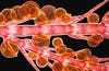
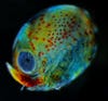
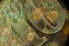
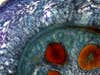
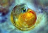
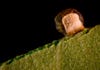
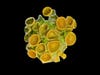
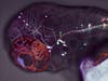
4th Place: Anglerfish ovary (4x)

This cross-section of an anglerfish’s ovary is reminiscent of another sea creature—the nautilus. Its otherwise-dull spirally composition caught James Hayden’s eye, so he used fluorescent hematoxylin and Eosin stains to illuminate the different details of the image. “I was trying to create an image that sharply defined the boundaries of the different parts of the specimen, so that the image could actually be used to demonstrate the morphology of the ovary and eggs,” he says.
This cute little guy is a juvenile seastar who just went through metamorphosis. It was likely accidentally caught by researchers netting plankton, and happened to transform in captivity. In the wild, the free-roaming larva's maturation would have necessitated a move to the bottom of the sea where it would have spent the rest of its life.

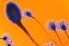
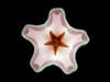
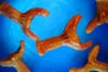
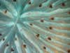
5th Place: Oral surface of a young seastar (40x)

This cute little guy is a juvenile seastar who just went through metamorphosis. It was likely accidentally caught by researchers netting plankton, and happened to transform in captivity. In the wild, the free-roaming larva’s maturation would have necessitated a move to the bottom of the sea where it would have spent the rest of its life.
Originating in the Amazon, the discus fish is becoming an increasingly popular aquarium fish due to the ease of its care. Known for their striking colors, discus are considered to fall into one of three species – green, red or blue/brown. Hormones and food coloring are sometimes used by sellers to enhance the color of these fish. Not only will these dyes fade over time, they can also do serious harm to the fish.

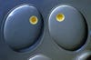
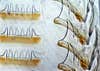
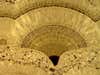
6th Place: Discus fish scales (20x)

Originating in the Amazon, the discus fish is becoming an increasingly popular aquarium fish due to the ease of its care. Known for their striking colors, discus are considered to fall into one of three species – green, red or blue/brown. Hormones and food coloring are sometimes used by sellers to enhance the color of these fish. Not only will these dyes fade over time, they can also do serious harm to the fish.
The friendly black-eyed Susan vine can be found in late summer and fall gardens all across America. This picture provides a look at its trichomes, hair-like epidermal cells reflect solar radiation, reducing internal temperatures and a plant's water loss. Interestingly, THC production takes place in the cannabis plant's trichomes. Although <em>Thunbergia alatas</em> don't offer that, they still look pretty darn cool.

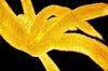
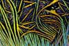
7th Place: Hair-like trichomes on Thunbergia alata (Black-eyed Susan vine) (450x)

The friendly black-eyed Susan vine can be found in late summer and fall gardens all across America. This picture provides a look at its trichomes, hair-like epidermal cells reflect solar radiation, reducing internal temperatures and a plant’s water loss. Interestingly, THC production takes place in the cannabis plant’s trichomes. Although Thunbergia alatas don’t offer that, they still look pretty darn cool.
These candy-colored cotton strands not only look pretty—they serve a purpose, too! Dr. Lloyd Donaldson snapped this shot of the stringy bits for a book on fiber structures. The use of confocal microscopy makes it possible to render 3-D images of tiny structures and then measure the diameter, for example, each thread.

8th Place: Cotton fibers stained with berberine sulphate and color depth shaded (200x)

These candy-colored cotton strands not only look pretty—they serve a purpose, too! Dr. Lloyd Donaldson snapped this shot of the stringy bits for a book on fiber structures. The use of confocal microscopy makes it possible to render 3-D images of tiny structures and then measure the diameter, for example, each thread.
Gabbro is an intrusive igneous rock that forms in molten hot magma chambers far below the earth's surface. Humans can see its coarse grains, usually black, with the naked eye but, in this case, Dr. Bernardo Cesare used a microscope to see even more. He says "an unusual abundance of small inclusions in the larger crystals" of this sample give it the shocking colors you see here.

9th Place: Olivine inclusions in gabbro (magmatic rock) (5x)

Gabbro is an intrusive igneous rock that forms in molten hot magma chambers far below the earth’s surface. Humans can see its coarse grains, usually black, with the naked eye but, in this case, Dr. Bernardo Cesare used a microscope to see even more. He says “an unusual abundance of small inclusions in the larger crystals” of this sample give it the shocking colors you see here.
Algae are often thought of as the pesky green gunk covering undisturbed ponds and lakes. What you might not realize, though, is the role algae and diatoms (which are actually oceanic plankton themselves) play in balancing the earth's climate. History has shown that the two contribute to the removal of billions of tons of carbon dioxide every year. Dr. Arlene Wechezak says she chose this photograph to "bring public awareness of the complexity and beauty of marine organisms."

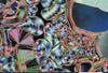
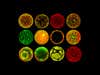
10th Place: Algae and diatoms (10x)

Algae are often thought of as the pesky green gunk covering undisturbed ponds and lakes. What you might not realize, though, is the role algae and diatoms (which are actually oceanic plankton themselves) play in balancing the earth’s climate. History has shown that the two contribute to the removal of billions of tons of carbon dioxide every year. Dr. Arlene Wechezak says she chose this photograph to “bring public awareness of the complexity and beauty of marine organisms.”
A translucent zebrafish larva showing the nervous system of the fish (green), the expression of the protein Tau (in red), and Alzheimer-like alterations of Tau (in blue). In humans Tau protein aggregates in the brains of Alzheimer patients and plays a major role in the degeneration of brain cells. By bringing Tau into translucent zebrafish larvae, the disease-causing effects of the protein can be studied directly under the microscope.

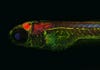
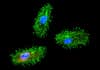
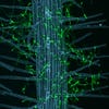
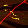
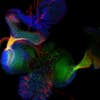
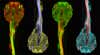
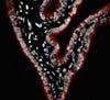
11th Place: “Alzheimer” Zebrafish, stained for Tau (red), neurons (green), and pathologic Tau (blue) (10x)

A translucent zebrafish larva showing the nervous system of the fish (green), the expression of the protein Tau (in red), and Alzheimer-like alterations of Tau (in blue). In humans Tau protein aggregates in the brains of Alzheimer patients and plays a major role in the degeneration of brain cells. By bringing Tau into translucent zebrafish larvae, the disease-causing effects of the protein can be studied directly under the microscope.
This piece of surreal art shows the flow patterns in a thin film of soap. A glass cylinder enclosed the flat soap film and kept it stable so that a close-up photograph could be taken. The striking black pattern is fringed with numerous pearl-like beads, a result of the fluid dynamics of the film.

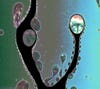
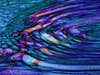
12th Place: Flow pattern in draining soap film (10x)

This piece of surreal art shows the flow patterns in a thin film of soap. A glass cylinder enclosed the flat soap film and kept it stable so that a close-up photograph could be taken. The striking black pattern is fringed with numerous pearl-like beads, a result of the fluid dynamics of the film.
Simple chemistry creates an otherworldly structure in this image. An organic solvent, an antiseptic and an outdated painkiller were combined to form this weird and complex crystal, illuminated with polarized light.

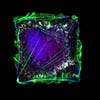
13th Place: Recrystallized melted mixture of acetanalide, resorcinal and carbon tetrabromide (33x)

Simple chemistry creates an otherworldly structure in this image. An organic solvent, an antiseptic and an outdated painkiller were combined to form this weird and complex crystal, illuminated with polarized light.
Nature's artistry is on display in this picture of a lobster egg a few days before hatching. The image was taken while scientists measured eye diameter, which is done to predict hatching time. The photography and microscopy may not have been too fancy, but the patterns and colors certainly are.

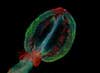
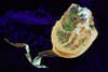
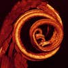
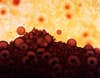
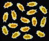
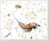
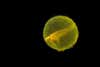
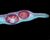
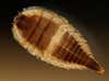
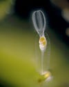
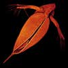
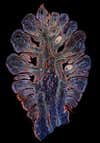
14th Place: Lobster egg (3.2x)

Nature’s artistry is on display in this picture of a lobster egg a few days before hatching. The image was taken while scientists measured eye diameter, which is done to predict hatching time. The photography and microscopy may not have been too fancy, but the patterns and colors certainly are.
What seems to be the head of some strange horned creature is actually the back end of a watersnipe fly larva. These weirdly shaped larvae are found in freshwater, and they and other bugs are often good indicators of water quality.

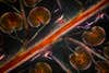
15th Place: Atherix ibis (fly) aquatic larva (25x)

What seems to be the head of some strange horned creature is actually the back end of a watersnipe fly larva. These weirdly shaped larvae are found in freshwater, and they and other bugs are often good indicators of water quality.
These symmetric ovals are actually the eggs of a pond snail. Snails deposits their eggs in large jelly-like clumps into the water. This picture shows a close-up of two of these eggs, with embryos developing inside them.

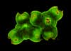
16th Place: Snail eggs (200x)

These symmetric ovals are actually the eggs of a pond snail. Snails deposits their eggs in large jelly-like clumps into the water. This picture shows a close-up of two of these eggs, with embryos developing inside them.
A colorful image showing the inner gears of a pocket stop watch. A confocal microscope, usually used to examine tiny biological materials, was used for this unusual look at the inner workings of the timepiece. This kind of microscopy can show the depth of the stop watch's various parts.

17th Place: Stopwatch (2.5x)

A colorful image showing the inner gears of a pocket stop watch. A confocal microscope, usually used to examine tiny biological materials, was used for this unusual look at the inner workings of the timepiece. This kind of microscopy can show the depth of the stop watch’s various parts.
This image captures a moment in the life of a human skin cell after it has been stimulated with a soluble growth factor. The cell shape was constrained, and after it was stimulated migratory structures extended from its membrane. The colors indicate proteins responsible for cell movement. By looking at how these proteins co-localize, scientists can understand how, when, and where they interact.

18th Place: Human skin on fibronectin with growth factor (60x)

This image captures a moment in the life of a human skin cell after it has been stimulated with a soluble growth factor. The cell shape was constrained, and after it was stimulated migratory structures extended from its membrane. The colors indicate proteins responsible for cell movement. By looking at how these proteins co-localize, scientists can understand how, when, and where they interact.
A photograph captures this rare type of snowflake. It only lasted for 2 hours out of three days of snowfall in Beijing, but at least the image endures.

19th Place: Snowflake (40x)

A photograph captures this rare type of snowflake. It only lasted for 2 hours out of three days of snowfall in Beijing, but at least the image endures.
The rust and dirt on this old coin transforms into a greenish-brown moonscape under the microscope. Found buried under the sand, the coin may have newfound value as a piece of abstract art.

20th Place: Rusted old coin (40x)

The rust and dirt on this old coin transforms into a greenish-brown moonscape under the microscope. Found buried under the sand, the coin may have newfound value as a piece of abstract art.
Gallery: This Year&#8217;s Most Amazing Microscopic Photography

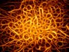
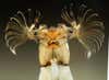
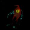
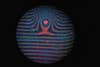
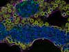
1st Place, Popular Vote: Fluorescent actin bundles growing from the surface of coated beads (63X)

Gallery: This Year&#8217;s Most Amazing Microscopic Photography

Honorable Mention: Ciliated protozoa (1700x)

Gallery: This Year&#8217;s Most Amazing Microscopic Photography

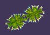
Honorable Mention: Fungal infection of Arabidopsis (flowering plant) root (25x)

Gallery: This Year&#8217;s Most Amazing Microscopic Photography

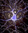
Honorable Mention: Rat cerebellum (200x)

Gallery: This Year&#8217;s Most Amazing Microscopic Photography

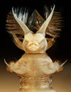
Honorable Mention: Whole finch testicle (4x)

Gallery: This Year&#8217;s Most Amazing Microscopic Photography

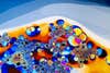
Honorable Mention: Fire agate (10x)

Gallery: This Year&#8217;s Most Amazing Microscopic Photography

Honorable Mention: Aspergillus mold in a microfluidic device (20x)

Gallery: This Year&#8217;s Most Amazing Microscopic Photography

Honorable Mention: Aspergillus sp. (250x)

Gallery: This Year&#8217;s Most Amazing Microscopic Photography

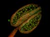
Honorable Mention: Arabidopsis thaliana (thale cress) anther (20x)

Gallery: This Year&#8217;s Most Amazing Microscopic Photography

Honorable Mention: Radula of Buccinum undatum (sea snail) (100x)

Gallery: This Year&#8217;s Most Amazing Microscopic Photography

Honorable Mention: Biosensing liquid crystals (20x)

Gallery: This Year&#8217;s Most Amazing Microscopic Photography

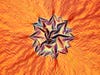
Honorable Mention: Hoya carnosa (wax plant) flower (10x)

Gallery: This Year&#8217;s Most Amazing Microscopic Photography

Honorable Mention: Pluteus larva of a sea biscuit (echinoderm) (200x)

Gallery: This Year&#8217;s Most Amazing Microscopic Photography

Honorable Mention: Water droplets ejected from a vibrating glass nozzle (200x)

Gallery: This Year&#8217;s Most Amazing Microscopic Photography

Honorable Mention: A fruit fly ovariole containing different stages of developing egg chambers (400x)

Gallery: This Year&#8217;s Most Amazing Microscopic Photography

Honorable Mention: Mosquito larvae (100x)

Gallery: This Year&#8217;s Most Amazing Microscopic Photography

Eriophorum vaginatum (Tussock cottongrass) corm (10X)

Gallery: This Year&#8217;s Most Amazing Microscopic Photography

Eriophorum vaginatum (Tussock cottongrass) corm (10X)

Gallery: This Year&#8217;s Most Amazing Microscopic Photography

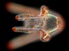
Moth proboscis (10X)

Gallery: This Year&#8217;s Most Amazing Microscopic Photography

2 pinnae of Aspidium Felix (fern) (20X)

Gallery: This Year&#8217;s Most Amazing Microscopic Photography

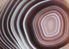
Agate – thin polished section (200X)

Gallery: This Year&#8217;s Most Amazing Microscopic Photography

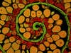
Branching filamentous diatom colonies with diatom ‘eyes’ (4X)

Gallery: This Year&#8217;s Most Amazing Microscopic Photography

Patterned expression of wild-type and transgenic Drosophila melanogaster (fruit fly) embryos (200X)

Gallery: This Year&#8217;s Most Amazing Microscopic Photography

Soap film (10X)

Gallery: This Year&#8217;s Most Amazing Microscopic Photography

Dover Sole skin (20X)

Gallery: This Year&#8217;s Most Amazing Microscopic Photography

Arabidopsis thaliana (thale cress) leaf epidermis and stomata (plant pores) (20X)

Gallery: This Year&#8217;s Most Amazing Microscopic Photography

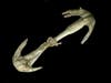
Antique microscope slide featuring Sea Cucumber skin and anchors (7.5X)

Gallery: This Year&#8217;s Most Amazing Microscopic Photography

Pyroxene and plagioclase (minerals) in granulite (metamorphic rock) (5X)

Gallery: This Year&#8217;s Most Amazing Microscopic Photography

Neurons extending from human embryonic stem cell spheres (20X)

Gallery: This Year&#8217;s Most Amazing Microscopic Photography

Grey cast iron (1000X)

Gallery: This Year&#8217;s Most Amazing Microscopic Photography

Re-crystallized urea

Gallery: This Year&#8217;s Most Amazing Microscopic Photography

Neuromuscular synapses in a Drosophila larva (200X)

Gallery: This Year&#8217;s Most Amazing Microscopic Photography

Diatom (480X)

Gallery: This Year&#8217;s Most Amazing Microscopic Photography

Embryo of guppy fish (40X)

Gallery: This Year&#8217;s Most Amazing Microscopic Photography

Two Micrasterias sp. (green algae), live specimens (200X)

Gallery: This Year&#8217;s Most Amazing Microscopic Photography

Two Closterium sp. (algae), live specimens (200X)

Gallery: This Year&#8217;s Most Amazing Microscopic Photography

Marine diatoms (200X)

Gallery: This Year&#8217;s Most Amazing Microscopic Photography

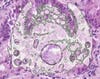
Bile duct surrounded by scar tissue in a cirrhotic liver (20X)

Gallery: This Year&#8217;s Most Amazing Microscopic Photography

Chromosome from Drosophila melanogaster (fruit fly) salivary glands (1000X)

Gallery: This Year&#8217;s Most Amazing Microscopic Photography

Benzoic Acid melt crystal (40X)

Gallery: This Year&#8217;s Most Amazing Microscopic Photography

Benzoic Acid melt crystal (40X)

Gallery: This Year&#8217;s Most Amazing Microscopic Photography

Echiniscus mediantus (tardigrade, water bear), in various states of movement

Gallery: This Year&#8217;s Most Amazing Microscopic Photography

Polished grey banded Carnelian (3X)

Gallery: This Year&#8217;s Most Amazing Microscopic Photography

Section of female Nippostrongylus brasiliensis (nematode) with eggs (200X)

Gallery: This Year&#8217;s Most Amazing Microscopic Photography

Mouse cranial base (200X)

Gallery: This Year&#8217;s Most Amazing Microscopic Photography

Human ileum (20X)

Gallery: This Year&#8217;s Most Amazing Microscopic Photography

A drop of pond water (100X)

Gallery: This Year&#8217;s Most Amazing Microscopic Photography

Gastric secreting cells of a mouse (40X)

Gallery: This Year&#8217;s Most Amazing Microscopic Photography

Axonal projections of an ommatidium of Drosophila eye (40X)

Gallery: This Year&#8217;s Most Amazing Microscopic Photography

Salamander larvae in egg with symbiotic green algae (1X)

Gallery: This Year&#8217;s Most Amazing Microscopic Photography

Green hydra on surface of spotted salamander egg mass (1X)

Gallery: This Year&#8217;s Most Amazing Microscopic Photography

Tilia sp. stem (500X)

Gallery: This Year&#8217;s Most Amazing Microscopic Photography

Silver sand (500X)

Gallery: This Year&#8217;s Most Amazing Microscopic Photography

Quercus leaf gall formed by a Gall wasp (4X)

Gallery: This Year&#8217;s Most Amazing Microscopic Photography

Gall formed by Trigonaspis mendesi (4X)

Gallery: This Year&#8217;s Most Amazing Microscopic Photography

Hippocampal neuron (63X)

Gallery: This Year&#8217;s Most Amazing Microscopic Photography

Transverse section of brown algae (40X)

Gallery: This Year&#8217;s Most Amazing Microscopic Photography

Crystal formed from desiccated phosphate-buffered saline (100X)

Gallery: This Year&#8217;s Most Amazing Microscopic Photography

Degenerating blue phase II crystals (100X)

Gallery: This Year&#8217;s Most Amazing Microscopic Photography

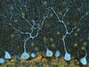
Mouse brain pyramidal cells (20X)

Gallery: This Year&#8217;s Most Amazing Microscopic Photography

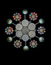
100-form exhibition diatom arrangement (200X)

Gallery: This Year&#8217;s Most Amazing Microscopic Photography

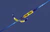
Elmis sp. (beetle) aquatic larva (60X)

Gallery: This Year&#8217;s Most Amazing Microscopic Photography

Head of Simulidae (fly) aquatic larva (40X)

Gallery: This Year&#8217;s Most Amazing Microscopic Photography

Natural textures of the SmF phase of 1,4-di-(n-tridecylthophene-2-yl)-benzene (200X)

Gallery: This Year&#8217;s Most Amazing Microscopic Photography

Dinosaur bone, Jurassic period (15X)

Gallery: This Year&#8217;s Most Amazing Microscopic Photography

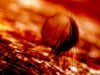
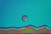
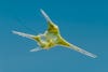
Raindrop on butterfly wing (20X)

Gallery: This Year&#8217;s Most Amazing Microscopic Photography

Lime tree leaf vessels (30X)

Gallery: This Year&#8217;s Most Amazing Microscopic Photography

Neuronally differentiated P19 cells (400X)

Gallery: This Year&#8217;s Most Amazing Microscopic Photography

Photonic crystals prepared from dried colloidal silica (5X)

Gallery: This Year&#8217;s Most Amazing Microscopic Photography

Salicornia europea (sprout) (20X)

Gallery: This Year&#8217;s Most Amazing Microscopic Photography

Urea and mannitol (100X)

Gallery: This Year&#8217;s Most Amazing Microscopic Photography

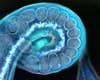
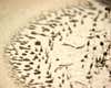
Stephanoceros fimbriata (rotifer) on a moss leaf (80X)

Gallery: This Year&#8217;s Most Amazing Microscopic Photography

Chalcedony with quartz in quartzite

Gallery: This Year&#8217;s Most Amazing Microscopic Photography

Chaetopleura Apiculata (Chiton) radula (100X)

Gallery: This Year&#8217;s Most Amazing Microscopic Photography

GaAs sample oxidized in water vapor (200X)

Gallery: This Year&#8217;s Most Amazing Microscopic Photography

Recrystallized melt mix of carbon tetra-bromide and resorcinal (33X)

Gallery: This Year&#8217;s Most Amazing Microscopic Photography

Forelimb bone from a rat

Gallery: This Year&#8217;s Most Amazing Microscopic Photography

Distribution of actin (green) & microtubule (red) cytoskeletons in fission yeast during mitosis (160X)

Gallery: This Year&#8217;s Most Amazing Microscopic Photography

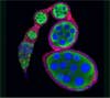
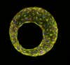
Life cycle of the social amoebae Dictyostelium discoideum (100X)

Gallery: This Year&#8217;s Most Amazing Microscopic Photography

Mouse Purkinje (brain) cells (40X)

Gallery: This Year&#8217;s Most Amazing Microscopic Photography

Dumbbell sponge spicule from an unidentified sponge (125X)

Gallery: This Year&#8217;s Most Amazing Microscopic Photography

Lily anther, transverse section, showing pollen sacs (100X)

Gallery: This Year&#8217;s Most Amazing Microscopic Photography

Lichen (10X)

Gallery: This Year&#8217;s Most Amazing Microscopic Photography

Notonecta Glauca (Backswimmer aquatic insect) (100X)

Gallery: This Year&#8217;s Most Amazing Microscopic Photography

Ceratium hirundinella (freshwater dinoflagellate), living specimen (400X)

Gallery: This Year&#8217;s Most Amazing Microscopic Photography

Filinia terminalis (rotifer) (250X)

Gallery: This Year&#8217;s Most Amazing Microscopic Photography

Microalgae in silicone matrix (1000X)

Gallery: This Year&#8217;s Most Amazing Microscopic Photography

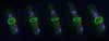
Zebrafish embryo, 22 hours post-fertilization, living specimen (40X)

Gallery: This Year&#8217;s Most Amazing Microscopic Photography

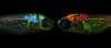
Drosophila melanogaster immune cells (200X)

Gallery: This Year&#8217;s Most Amazing Microscopic Photography

Three day old zebrafish larva with fluorescently labeled neurons (20X)

Gallery: This Year&#8217;s Most Amazing Microscopic Photography

Microprocessor with a pollen grain (50X)

Gallery: This Year&#8217;s Most Amazing Microscopic Photography

Mouse cultured dorsal root ganglion neuron and satellite cells (1200X)

Gallery: This Year&#8217;s Most Amazing Microscopic Photography

Xenopus laevis (African clawed frog) tadpole (10X)

Gallery: This Year&#8217;s Most Amazing Microscopic Photography

Ammonium Dichromate, crystallized (160X)

Gallery: This Year&#8217;s Most Amazing Microscopic Photography

Beer bubbles (160X)

Gallery: This Year&#8217;s Most Amazing Microscopic Photography

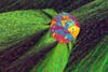
Oenothera biennis (Evening primrose) pollen grains (650X)

Gallery: This Year&#8217;s Most Amazing Microscopic Photography

Top of metal tufting hook used in a tufting machine to make carpets (5X)

Gallery: This Year&#8217;s Most Amazing Microscopic Photography

Antique microscope slide featuring arranged diatoms (60X)

Gallery: This Year&#8217;s Most Amazing Microscopic Photography

Fish scale base, whole mount (125X)

Gallery: This Year&#8217;s Most Amazing Microscopic Photography

Bundles of quartz fibers with linking bands of Fe-hydroxide (10X)

Gallery: This Year&#8217;s Most Amazing Microscopic Photography

Dennstaedtia sp. (cup fern) longitudinal rhizome section (10X)

Gallery: This Year&#8217;s Most Amazing Microscopic Photography

Photonic crystal structure manufactured holographically (100X)

Gallery: This Year&#8217;s Most Amazing Microscopic Photography

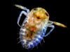
Ventral view of Daphnia pulex (common water flea) (10X)
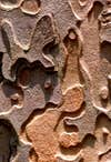
Gallery: This Year&#8217;s Most Amazing Microscopic Photography

Pine skin (10X)

Gallery: This Year&#8217;s Most Amazing Microscopic Photography

Alzheimer Zebrafish, stained for Tau (red), neurons (green), synapses and pathologic Tau (blue) (10X)

Gallery: This Year&#8217;s Most Amazing Microscopic Photography

Rat kidney epithelial cells on a fibronectin surface pattern (630X)

Gallery: This Year&#8217;s Most Amazing Microscopic Photography

Giant liposomes of pulmonary surfactant (40X)

Gallery: This Year&#8217;s Most Amazing Microscopic Photography

Arabidopsis thaliana (thale cress) stigma (20X)

Gallery: This Year&#8217;s Most Amazing Microscopic Photography

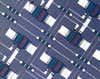
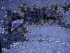
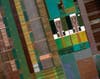
Section of circuit board (20X)

Gallery: This Year&#8217;s Most Amazing Microscopic Photography

Mullerian epithelium from human fallopian tube (400X)

Gallery: This Year&#8217;s Most Amazing Microscopic Photography

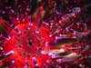
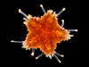
Echinometra lucunter (sea urchin), oral view (20X)

Gallery: This Year&#8217;s Most Amazing Microscopic Photography

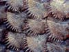
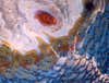
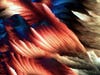
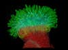
Coral, live polyp showing its mouth, glowing tissue and brown tentacles covering the stony skeleton (6X)

Gallery: This Year&#8217;s Most Amazing Microscopic Photography

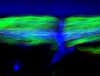
Neuropil in a zebrafish epithalamus (40X)

Gallery: This Year&#8217;s Most Amazing Microscopic Photography

Pinus Ovulate cone mother cell (10X)

Gallery: This Year&#8217;s Most Amazing Microscopic Photography

Carcinoma cells (40X)

Gallery: This Year&#8217;s Most Amazing Microscopic Photography

Dried toluidine blue on a Petri dish (35X)
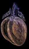
Gallery: This Year&#8217;s Most Amazing Microscopic Photography

Mammal heart (10X)