The Ultimate Operating Room
If you happen to need brain surgery, the place to be is M.D. Anderson Cancer Center at the University of Texas in Houston. The center’s new $9.2-million operating room, called the BrainSUITE, features one of the most sophisticated neurosurgical setups on the planet (soon to be available in seven other U.S. hospitals). Ceiling-mounted cameras give surgeons a magnified view of the brain, giant “data billboards” display vital signs throughout the room, and an extra-wide MRI scanner can accommodate bodies in nearly any position, so patients lying on their side don’t need to be bandaged up and rolled onto their back to slide into the machine. A surgeon simply swivels the operating table into the scanner to inspect his handiwork. (Scalpels and other magnetic objects, however, must first be moved out of range of the magnet.)
All of this helps to eliminate the dangerous guesswork inherent to brain surgery. With brain tumors, for instance, a surgeon must remove 98 percent of the growth to give the patient a shot at surviving beyond one year. This creates a delicate balancing act. Take out too much tissue, and you increase the chance of damaging other parts of the brain. But leave behind more than two percent of the mass, and it might regenerate, shaving as many as four months off the patient’s lifespan. With the MRI-equipped BrainSUITE, a surgeon can determine in a matter of minutes whether there’s a need to go back in to finish the job-and can get the patient off the table as quickly and safely as possible.-Michael Rosenwald
For a look at laser scalpels, skull drills, and robotic arms on hand to aid today’s neurosurgeons, launch the gallery here.
The No-Harm Robo-Arm
Sticking your head in an MRI scanner while robotic hands cut into your brain may sound like a terrifying proposition–but at least they won’t accidentally nick your optic nerve. After five years and $27 million spent, the University of Calgary and the Calgary Health Region health-care system have unveiled the neuroArm, the first surgical robot that operates inside an MRI scanner, combining high-precision surgery with near real-time imaging. Most surgeons operate within one- or two-millimeter accuracy, says project leader Garnette Sutherland. The neuroArm can make incisions accurate to 50 microns–about half the diameter of a human hair. A surgeon using the neuroArm doesn’t even need to be in the same room as the patient–a pair of high-definition cameras mounted on a microscope streams images to the doctor’s control station. The controls filter out hand tremors, while a haptics system provides tactile feedback. The haptics can also help prevent accidental damage. “I can set a zone between the optic nerve and carotid artery, and the machine won’t let me touch either of them,” Sutherland says. Pending a successful debut surgery–most likely a brain-tumor operation this fall–and FDA approval, Sutherland says the robot will reduce surgical error and HMO bureaucracy in one office visit. Typically a patient will undergo an MRI, come back for a consultation, and finally return for a biopsy. “Now,” Sutherland says, “if we see a lesion in MR imaging, we could biopsy it on the spot.”–Michael Slenske
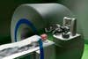
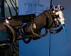
Robotic Arsenal
The neuroArm is outfitted with a cauterizer, 12 micro-dissectors, a suction hose, bayonet-shaped micro-scissors, and needle drivers to hold sutures.
Nurse, Pass The Femtolaser
Though he’s performed more than 8,000 brain surgeries in his 30-year career, Michael Apuzzo hates using a scalpel, a tool he considers woefully outdated. “Whenever I go in to do a surgery with a scalpel in my hand,” says Apuzzo, a professor of neurological surgery at the University of Southern California, “I feel like I should be wearing a powdered wig.” Soon Apuzzo may be able to swap his 18th-century cutting tool for a laser. Femtosecond lasers, the fastest in the world, are capable of producing energy pulses that last a millionth of a billionth of a second and can be focused into beams less than one hundredth the diameter of a human hair. This makes them ideal for operating on subcellular structures-such as the axon, the long tail by which a neuron sends information to its neighbors-that are far too small for even the finest robotic surgical hands to handle. Biologist Yishi Jin of the University of California at Santa Barbara and mechanical engineer Adela Ben-Yakar of the University of Texas led a team that used the femtolaser to sever the axons that control muscles in nematode worms. The worms immediately lost the ability to wiggle backward, although they regenerated about half their axons, and some worms regained full movement within 24 hours. The ability to operate on individual brain cells without killing them in the process could allow scientists to study how the cells regenerate and might lead to better treatments for neurodegenerative diseases such as Alzheimer’s and Parkinson’s. Someday, doctors could even use the laser to slice out damaged axons and replace them with new ones. With far-off applications such as these, the femtolaser has the potential to take neurosurgery way beyond the limits of its powder-wigged origins.-Andrew Rosenblum At left: The laser zaps the axon with quadrillions of pulses of light per second, creating a spot on the cell that is as hot as the sun. (The beam’s tight focus and short duration prevent heat from building up in surrounding cells.) The axon evaporates off [inset] as a plasma of electrons and ions, leaving behind scant debris.
Instant Feedback
Doctors can scan the patient’s brain multiple times during surgery, immediately revealing how much of a tumor remains to be removed.
Pulling Out Tumors Through Your Nose
Brain surgery involves actually cracking open skulls, which often leads to post-op complications and a lengthy recovery. But Amin Kassam, chairman of the department of neurological surgery at the University of Pittsburgh Medical Center and director of its Minimally Invasive Endoneurosurgery Center, has found a way to leave your melon intact: He performs brain surgery through the nose. Kassam, who helped pioneer the technique in the late 1990s, enters the nostril and goes into the sinus cavity, where he drills a small “keyhole” into the base of the skull. He then carefully peels away the three membranes that encase the brain to access the entire bottom half of the organ, a region that extends between the forehead to the top of the spine and from ear to ear. Using angled endoscopes he designed himself and guided by a GPS-like system that tracks the scopes and maps the brain in near real time, Kassam cuts up and suctions out tumors or repairs aneurysms in an otherwise conventional manner. He then patches the keyhole with a flap of tissue from the nostril, leaving no visible scars and only a minor incision to heal. In many cases, patients are back on their feet in a matter of days, instead of weeks. To date, Kassam has performed more than 750 procedures (in one notable case, he removed a baseball-size tumor from a young boy’s head) and teaches the technique to surgeons every year. He is one of only a handful of surgeons in the U.S. who perform the surgery, but he thinks that number will grow as more surgeons realize the benefits. “As long as the important stuff-like nerves and major blood vessels-is on the outside,” he says, “I’ll come from the inside.”-Jason Daley At left: By going through the nose, what were once incredibly invasive procedures now leave no visible scar.
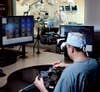
Hand-Eye Coordination
An array of monitors helps surgeons keep an eye on progress and reference pre-op scans as they use a joystick and stylus to control the neuroArm’s robotic hands.
The World’s Most Sensitive Robotic Drill
When cutting into a person’s brain, the slightest change in tissue density can go unnoticed by even the most experienced hands. But help could soon arrive in the form of a hypersensitive robotic drill that stops drilling through bone as soon as it reaches the soft stuff on the other side. Peter Brett, a biomedical engineer at Aston University in England, built the drill with three sensors that detect when the tip pierces different types of tissue. The drill measures the pressure and twisting forces it comes into contact with at each layer to identify the material and predict how it will react to being pierced. For instance, a sudden drop in pushing force and a spike in torque signals that the drill has reached the delicate fibrous membrane that encases the brain. A third sensor monitors the tissue’s flexibility to determine how much material remains to be drilled through. The robotic micro-drill beats the human competition in both cleanliness and reaction time. Traditional surgical drills bore beyond their target depth, leaving bony debris in their wake that can contaminate and damage the brain. In contrast, Brett’s device stops and reverses course at the first touch of membrane, allowing surgeons to clear skull fragments before going deeper. In April, David Proops, a surgeon at the University Hospital Birmingham NHS Foundation Trust in England, used the drill to bore holes less than a millimeter wide into the cochleae of three hard-of-hearing patients. Brett suggests that the fine-tuned power tool could be used to secure pins into the hard bone tissue of brain-injury victims, and he’s currently investigating funding options for the instrument’s federal approval processes in the U.K. and the U.S. If all goes well, within three years the drill will be helping doctors perform more sensitive surgeries.-Corey Binns At left: The robotic microdrill is so sensitive that it can bore through the shell of an egg and stop before rupturing the thin membrane that encases the albumen.
![Though he's performed more than 8,000 brain surgeries in his 30-year career, Michael Apuzzo hates using a scalpel, a tool he considers woefully outdated. "Whenever I go in to do a surgery with a scalpel in my hand," says Apuzzo, a professor of neurological surgery at the University of Southern California, "I feel like I should be wearing a powdered wig." Soon Apuzzo may be able to swap his 18th-century cutting tool for a laser. Femtosecond lasers, the fastest in the world, are capable of producing energy pulses that last a millionth of a billionth of a second and can be focused into beams less than one hundredth the diameter of a human hair. This makes them ideal for operating on subcellular structures-such as the axon, the long tail by which a neuron sends information to its neighbors-that are far too small for even the finest robotic surgical hands to handle. Biologist Yishi Jin of the University of California at Santa Barbara and mechanical engineer Adela Ben-Yakar of the University of Texas led a team that used the femtolaser to sever the axons that control muscles in nematode worms. The worms immediately lost the ability to wiggle backward, although they regenerated about half their axons, and some worms regained full movement within 24 hours. The ability to operate on individual brain cells without killing them in the process could allow scientists to study how the cells regenerate and might lead to better treatments for neurodegenerative diseases such as Alzheimer's and Parkinson's. Someday, doctors could even use the laser to slice out damaged axons and replace them with new ones. With far-off applications such as these, the femtolaser has the potential to take neurosurgery way beyond the limits of its powder-wigged origins.-Andrew Rosenblum <em>At left:</em> The laser zaps the axon with quadrillions of pulses of light per second, creating a spot on the cell that is as hot as the sun. (The beam's tight focus and short duration prevent heat from building up in surrounding cells.) The axon evaporates off [inset] as a plasma of electrons and ions, leaving behind scant debris.](https://www.popsci.com/uploads/2019/03/18/DCH7OUNSG4VGZYZ5HEGW47EJZ4.jpg?auto=webp&optimize=high&width=100)