Each year, the Wellcome Trust in the UK hosts an image competition that showcases medical science at work. The awards honor the “most informative, striking and technically excellent images” added to Wellcome’s impressive collection.
The entire gallery is on display in the UK and on the Wellcome website, but we have a sampling for you here.
_
Click to launch the photo gallery_
Caffeine Crystal
This false-color scanning electron micrograph shows caffeine crystals. In plants, caffeine functions as a natural pesticide that paralyzes and kills some insects. The main crystals were 400-500 microns long; however, this crystal group formed on the end of the larger crystal and measures around 40 microns in length. The judges selected this image because it’s such an unfamiliar perspective: “It’s a bright, intricate image of something that most of us experience every day.”
Arabidopsis Thaliana Seedling
This confocal micrograph shows the tissue structures within the leaf of an Arabidopsis thaliana seedling, the model organism in plant genetics. The sample was fixed and stained with propidium iodide, which labels DNA, but was imaged four years later. Over time, oxidation of the stain in different parts of the tissue provides different fluorescent properties, which researchers are using to study plant cellular architecture and gene activity.
Frog Eggs
… Xenopus laevis oocytes, to be exact. Otherwise known as the African clawed frog, this is another model organism used in developmental biology research. Each oocyte is surrounded by thousands of follicle cells, shown by staining DNA blue. Blood vessels, which provide oxygen to the oocyte and follicle cells, are shown in red.
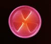
Radioactive Symbol
This diatom frustule is a type of phytoplankton. The organisms are encased inside hard cell walls, and come in a variety of shapes, pores and ridges. These features are used to determine their genera and species. This diatom is 80 microns in diameter and just happens to look a lot like the universal symbol for radioactivity.
Beautiful Biofilm
This confocal micrograph shows Bacillus subtilis, a large rod-shaped bacteria commonly found in soil. This is a biofilm showing distinct lineages of the microbe, which were stained with different fluorescent proteins and then mixed randomly on a petri dish. As they grow, they organize into reproducible patterns and shapes that can be predicted with mathematical models.
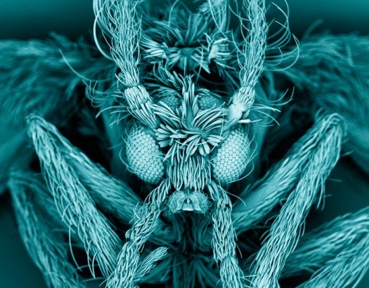
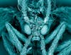
Moth Fly
This false-color scanning electron micrograph shows a moth fly, Psychodidae, also known as a drain fly. The larvae live in domestic drains and emerge in sinks. Their bodies and wings are covered in fine cilia, which lend them a moth-like appearance.
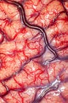
Overall Winner: Brain Surgery
This photograph shows the cortex of a human brain belonging to an epileptic patient. It was taken before an intracranial electrode procedure to treat the patient’s epilepsy. After removal of several brain sections, this patient made a full recovery and no longer suffers from seizures. Judges chose it as the overall winner because of its detail. From judge Alice Robert explains: “The ‘gray’ matter (which is gray in death) is blushing pink. Small arteries are glowing with the scarlet blood pulsing through them, while purple veins lie thickly in the sulci, the crevices of the brain. And underneath that is somebody’s mind.” See the other winners in the full gallery over at the Wellcome Trust.