Oversize Sculptures Offer a Close Look at Bacteria and Viruses
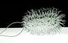
This 41-inch-long sculpture of the Escherichia coli bacterium is part of British artist Luke Jerram’s “Glass Microbiology” series of portraits....
Seen Clearly
This 41-inch-long sculpture of the Escherichia coli bacterium is part of British artist Luke Jerram’s “Glass Microbiology” series of portraits. Other organisms he has vitrified include HIV, SARS and swine flu.
To create each one, Jerram used images from an electron microscope and had guidance from virologist Andrew Davidson of the University of Bristol in England. “Scientists have to jump from what they can see [in the microscope] to what they know through chemical analysis, and then they have to piece together a kind of jigsaw,” Jerram says. He takes the scientists’ microscopy data and analysis a step further, transforming diagrams and images into three-dimensional models. But why glass? The color-blind artist wished to challenge the mainstream media’s love for artificially colored images of these minuscule attackers by rendering the organisms in a less fanciful palette.