How a kickball helped surgeons heal a fetus
Before operating within a living womb, they had to practice.
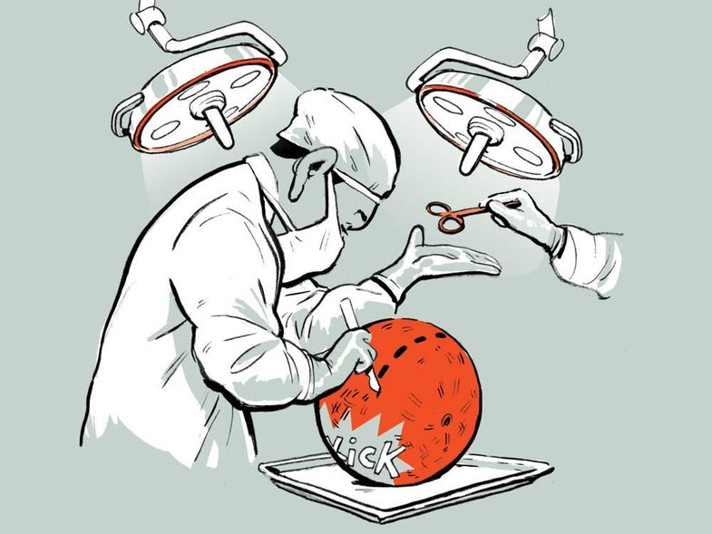
↑ Oluyinka Olutoye, pediatric surgeon and co-director of the Texas Children’s Fetal Center
I am a pediatric surgeon, but I meet many of my patients when they’re still growing inside their mother’s womb. That’s because I specialize in fetal surgery—performing an operation on a baby before it’s born.
Usually, we remove the baby from the uterus, operate, then put it back. But you can’t always do that. For example, spina bifida, a defect where bone and flesh don’t fully cover the spinal cord, requires intervention at around 22 to 25 weeks of gestation. If we make a large cut in the uterus at that time, when the fetus weighs less than a pound, the organ could tear as the baby grows, endangering mother and child.
To get around that, doctors at my hospital created a technique to operate without opening the womb. We make two tiny incisions in the uterus. Then we insert probes with small cameras at the ends and carefully use them to repair the tissue over the exposed spinal cord. This limited access makes the surgery incredibly tricky, and the margin for error in a person that tiny is minuscule. So, before we operate on real babies, we practice with a simulation. We sew chicken skin to the back of a baby doll, in the exact location of the patient’s spinal defect, and place it inside a kickball “uterus.”
Chicken skin is a little tougher than fetal skin but far more similar to the real thing than plastic—and pretty easy to obtain at a grocery store. This lets us practice inserting the probes into the kickball and operating on the doll.
I once operated on a baby that I could have fit in the palm of my hand. Today, that child is alive and well—and far bigger. I ask each of the kids I treat, “Do you know how small you were when I first met you?” I tell them, and they always say, “No, that’s not possible.” But it is.
As told to Claire Maldarelli
This article was originally published in the Fall 2018 Tiny issue of Popular Science.
