Why Give a Dead Man a Body Scan?
Forensic scientists in Switzerland are pioneering a whole new way to do autopsies. No scalpel required.
A light shines under the closed door of a radiology suite, down a darkened hallway deep inside the University Medical Center in Bern, Switzerland. Outside the building, under the glow of a fluorescent street lamp, an empty hearse waits in the loading dock. Tonight the local undertaker is earning some extra money making a special delivery. Entering the radiology room through a back door, he gently deposits a body–double-wrapped inside a blue bag–on the sliding bed of a full-body scanner. The bag, through which x-rays can easily pass, will remain closed while the body is scanned, both to respect the privacy of the dead and so as not to disturb any nonforensic personnel in the room.
Without the bag, the university’s Institute of Diagnostic Radiology would not have approved the use of its aseptically clean research facilities for postmortem studies, says forensic pathologist Michael Thali. The Swiss emphasis on orderliness and precision extends to the task of death investigation.
This cultural passion–some would say obsession–for precision becomes clear to any visitor arriving by train here in Switzerland’s 800-year-old, meticulously preserved capital. Rows of clocks line the train station corridor, all perfectly synchronized down to the sweep of their prominent second hands. In this spirit, Thali and his colleagues at Bern’s Institute of Forensic Medicine are perfecting the ultimate no-mess autopsy: precise, objective and nondestructive, with death’s every data point captured permanently on compact discs that the scientists store in the vault of a nearby Swiss bank (where else?).
Thali calls the technique “virtopsy,” or virtual autopsy. Specifically, his research team has adapted the twin medical- imaging technologies of computed tomography (CT) and magnetic resonance imaging (MRI) to create three-dimensional, high-resolution computer images of a crime victim’s internal organs. Thali pours these digitized blood and guts into a hollow-man replica of the victim. The result is a head-to-toe cybercorpse that a pathologist can view–wounds and all–from any depth and angle, including inside out.
Besides being a bloodless approach to an otherwise messy job, the digitally preserved bodies of the Virtopsy Project have the added benefit of permanency. “Murder victims have the unfortunate habit of decomposing,” Thali notes. Of course, police and pathologists have long documented such disappearing evidence with photographs and detailed medical reports. Photos, however, are limited by their two-dimensionality and the inherent distortion of camera angles. And medical reports, according to Thali, remain unacceptably subjective.
It’s a criticism supported by the cacophony of the courtroom, where prosecutors and defense lawyers often present dueling pathologists, each reinterpreting autopsy reports to favor one side or the other. Complicating a jury’s difficulty in following such arguments are the typically gore-drenched autopsy photos that prompt many to turn away in horror. “We [in Switzerland] are not so used to shows like CSI,” Thali points out. “It can be a real problem.”
In the future that Thali envisions, any pathologist taking the witness stand can bloodlessly redissect the victim in full view of the jury by calling forth the original data stored on the discs. “Graphic, yes. Gory, no,” he says.
Over the past three years, Thali has performed more than 100 virtual autopsies, each followed by a traditional autopsy to confirm his findings. Although his experimental technique has proved highly accurate, he expects to complete at least 100 more cases before the first virtopsy debuts in a court of law.
“Virtopsy is still like a little baby,” Thali says. “It is not yet ready to stand alone.” First he must show that it is at least as accurate as traditional autopsy. So far, he says, virtopsy has been particularly good for detecting the internal bleeding, bullet paths and hidden fractures that can be maddeningly difficult to
isolate amid the mass of blood and gore that results when a pathologist is forced to essentially eviscerate the body.
Best of all, perhaps, is the way CT and MRI scans highlight emboli–air bubbles that obstruct blood vessels and that have most likely entered the body through a wound of some sort. Such effervescent evidence can vanish as soon as a pathologist slices open a vein or organ to look for it, Thali explains. “So difficult is this problem that some have proposed performing underwater autopsies in swimming pools to detect escaping air bubbles,” he says.
The scans also make it much easier to detect aspirated, or inhaled, water and blood in the lungs. These forensic “vital signs” tell a pathologist that a victim was alive when he entered the water or sustained an injury, which can be crucial for determining whether an apparent drowning or car crash was staged to cover up a murder. Pockets of air, blood or water show up clearly on CT and MRI scans as spots–black, bright white or gray–against the background of body tissues.
On the negative side, virtopsy remains woefully inadequate for diagnosing poisoning, as well as common natural causes of death such as infection or heart failure. “Obviously,” Thali admits, “it’s very important to be able to rule out such natural causes in a case of suspected murder.”
The forensic question before Thali tonight is whether or not the elderly woman inside the body bag was dead before she ended up under the chassis of a Volvo sports sedan the previous afternoon. The Volvo’s driver insists that he checked his rearview mirror before backing into the parking stall where the body was found. Given the woman’s age–70ish–the possibility of a prior heart attack or stroke seems plausible.
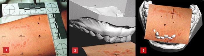
Thali’s research team began their examination of the body earlier in the day, as it lay face-up on the stone examining table in the forensic institute’s second-floor autopsy bay. Visualization specialist Ursula Buck and pathology resident Emin Aghayev prepared the body by affixing buttonlike reference markers across its surface and photographing it with a digital camera from nine angles. Using an overhead light and transparency, they then projected a numbered grid of black points across the body before initiating a computer-guided 3-D scan using cameras mounted on an overhead beam. Turning the body over, Buck and Aghayev repeated the procedure before placing the corpse in a bag and sending it, by private hearse, to the university’s Institute of Neuroradiology, several blocks away, for its MRI scan.
Magnetic resonance imaging has become fairly routine in medical diagnostics since its introduction in 1980. Using radio waves beamed through a powerful magnetic field, MRI produces 3-D internal images of unsurpassed detail. But the process remains far from automated, requiring operators to learn elaborate protocols to extract images from different types of body tissue. Complicating matters for the virtopsy project, MRI technologist Karin Zwygart has had to create special protocols to compensate for the lower body temperatures of Thali’s refrigerated research subjects. The cooler temperatures would otherwise wreak havoc on results, because the MRI machine operates by translating the signature vibrations emanating from the nuclei of different kinds of atoms. At cooler temperatures, these nuclear vibrations slow down.
On the plus side, the resulting images are of such clarity that they draw amazed inquiries from radiologists whenever Thali displays them at international conferences. “Not only do they not fidget,” he says of the corpses, “there is no beating heart, no circulating blood, no digestive motions to blur our images.”The body’s final appointment of the night brings it to the University of Bern’s Institute of Diagnostic Radiology. Here it passes through the doughnut-shaped hole of a CT scanner, which constructs 3-D images of the body from a series of x-ray slices. In the radiology suite’s darkened computer room, Peter Vock, director of the imaging institute, shares a computer with neuroradiologist Luca Remonda. As intent as schoolboys with a new videogame, the two men take turns clicking and dragging screen controls to manipulate the image on the monitor. Vock defines and deletes the CT scanner’s bed to leave the woman’s body suspended in midscreen. Slowly he melts away silvery layers of skin, muscle and connective tissue to reveal a bare white skeleton. He rotates the image, head over heels, pausing to note multiple rib fractures, a broken sternum, a shattered collarbone and crushed vertebrae. Then, layer by layer, he reassembles the body. When he reaches the level of fascia–midway between bare bones and full muscle–he stops again, intrigued by the abnormally high position of the woman’s stomach and the telltale indentation, like an overly tightened belt, around the organ’s midsection.
“We call this a collar sign,” Vock explains. “We think that perhaps the woman’s stomach was pushed up through a break in her diaphragm,” the large muscle that separates the lungs from the abdominal organs. To get a better look, Vock finishes reconstructing the torso and then slices down through its midsection, five millimeters at a click, until he discovers a dark gap in the white muscle of the diaphragm.
Vock turns the controls over to Remonda, and Thali asks the radiologist to look for signs of inhaled blood in the woman’s lungs. Earlier, during the MRI scan, Thali noted light areas in the woman’s muscle tissues. If this represented active bleeding, it would suggest that she was alive when the car crushed her body against the pavement. But postmortem injuries can produce some internal blood seepage. More telling would be a clear indication of aspirated blood–a confirmation that the woman was still breathing when she sustained the injuries.
Remonda is on his second or third slice through the lungs when the first white splotches appear. As he continues to click down through the tissues, each bright spot melts away, only to be replaced by others. Clearly, aspirated blood.
Remonda and Vock turn to Thali for his pronouncement as to probable cause of death. “Thorax instability secondary to crushing injuries,” he concludes: suffocation following rib fractures so massive that the victim was unable to take a breath.
The next morning Richard Dirnhofer, Thali’s boss and the director of the Institute of Forensic Medicine, will perform a conventional autopsy for confirmation. “It will be a bloody mess, to be sure,” Thali says with a grimace. “Nobody likes to show that to a jury.”At the same time that Dirnhofer is making his first cut, visualization specialist Buck will be hauling her surface-scanning equipment to the police garage where the suspect’s Volvo remains impounded. There she will create a 3-D computer image of the car’s surface, the same way she photographed the corpse. Buck and Thali will then bring the body and automobile together in cyberspace, matching wounds to car surfaces until they can determine the woman’s position as she sustained each of her injuries. Thali is particularly interested in how well the car’s rear bumper and trunk lid will match up against the gouges seen in the surface scan of the woman’s knees and the deep bleeding the MRI picked up in the muscles over her hips. “This is not some animation program using artificially created models,” Thali insists. “You are looking at real data from the original car, the original person, the original wounds and bones.” Depending on how the data match up, the driver could face manslaughter charges.
This dynamic matching of wound to weapon is an offshoot of a larger collaboration between Thali’s forensic pathology team in Bern and the Scientific Forensic Service of the Zurich police. In 1995 Bern’s Dirnhofer called Zurich’s chief of forensics, Walter Brschweiler, with a challenge. He wanted a better way to match wounds and suspected weapons than the usual method of laying a photo of one over a photo of the other.
“That is when I thought of Marcel,” Brschweiler says. Zurich traffic detective Marcel Braun had been adapting the new technology of photogrammetry for use in accident investigation. Photogrammetry software–originally developed for topographical mapmaking–turns a calibrated series of photographs into a 3-D model. In essence, Braun was using it to rewind multi-vehicle traffic accidents, extrapolating back from the dents and twisted metal of the final pileup to see who hit whom, all the way back to first impact. In collaboration with Thali, Braun adapted the technique to re-create violent deaths from the resulting damage seen on and within the corpses.
One of the most interesting cases on which the Bern and Zurich forensic scientists have collaborated came to trial in July 2003. It was a triple homicide, with three prostitutes found beaten to death in an apartment outside Zurich. Police had
a suspect who admitted to having sex with the women but insisted that they were all alive when he left the apartment.
The murder investigation focused on a deep bite mark gouged into the shoulder of one victim. The man denied biting anyone, and DNA swabs of the wound yielded a mixture of genetic fingerprints that would not stand up in court. The possibility remained that the women had killed each other. Indeed, judging from the size of the bite mark, police originally proposed that the bite on the woman’s shoulder came from one of the other two victims. And when the Zurich police obtained dental casts from the other women’s mouths, one did fit fairly well with their photographs of the wound.
Meanwhile Thali’s team had completed their virtopsy of the three women, with Braun performing the 3-D surface scans. They could now go beyond matching dental casts against two-dimensional photos, to re-create how the teeth penetrated the dead woman’s skin.
On a recent morning in his Zurich office, Brschweiler replays the results on his laptop computer–calling up the digitized, silvery-gray replica of the male suspect’s teeth and bringing it in contact with the full-color virtopsy scan of the victim’s bruised and bloodied shoulder. As the front teeth begin to enter the skin, Brschweiler switches to an underside view. “You see, there is not yet a reaction from the victim,” he
narrates, “only the motion of the biter.” But as the premolars break through, the teeth begin to drag laterally, widening the wound. “Now see, the woman reacts to the bite. She begins to pull away.”
Re-running the simulation again, even slower, Brschweiler underscores the perfect match between teeth and bite marks. He then calls up the digitized dental cast of the dead woman whom police originally linked to the bite. “It matches on the left, but the angle is not the same,” he says. “And look here, the small gap between the front teeth is not a perfect match.” Importantly, Brschweiler says, the judge and jury were able to follow the re-creations and the science behind them during the suspect’s murder trial, which resulted in a conviction.
In their shared pursuit of more-accurate murder re-creations, the Zurich Police Department and the Virtopsy Project have enlisted the additional help of the Swiss army, which has invested a considerable peace dividend in science. Switzerland has the distinction of ranking simultaneously among the world’s most peaceful and most militarized nations, having avoided war for more than 150 years while enlisting virtually every male citizen in its military reserves.
“We have the time, people and peace to spend time in research,” says Swiss Department of Defense mathematician Beat Kneubuehl, an internationally recognized expert on wound ballistics–the physical dynamics of how bullets, their fragments and their associated air jets pass through the human body. Kneubuehl’s interest in gunshot wounds dates back to 1978, when the Swiss army asked him to design a line of ammunition and demonstrate that it met international conventions against unnecessary suffering.
To prove that his bullets would pass through a leg or arm without causing the kind of massive bone and blood-vessel damage that results in amputation, Kneubuehl decided to use simulated body parts instead of cadavers or animal carcasses, partly for ethical reasons. “We Swiss frown on that sort of thing,” he says. But the major offense to Kneubuehl’s Swiss sensibilities was the imprecision of such targets. “Every cadaver, every human or animal bone is a little bit different from the next one,” he explains. “When I want to isolate one variable–the design of my bullet–I must expunge all other variables from my experiments.”
Kneubuehl’s line of faux body parts fracture, splatter, and shred like the real stuff–just more consistently so. His simulated bone consists of two layers of polyurethane sandwiching an inner layer of gelatin and coated with a thin veneer of rubber. His skulls are melon-shaped spheres with a corked hole for adding gelatinous brain and fake blood. They have been useful for re-
creating fatal beatings and shootings.
On one of Kneubuehl’s recent test days, Thali joins him at the Swiss army’s wooded training grounds outside the Alpine village of Thun. The sound of machine-gun fire alternates with that of birdsong as the two men enter the smallest of the site’s three underground ballistic-test tunnels. Built in the early 1990s to minimize noise disturbance to Thun residents, the 100-, 200- and 500-meter tunnels are the largest underground firing ranges in the world.
Kneubuehl’s goals for the day are to study the fleeting expansion of brain tissue caused by the air jet that accompanies a bullet and to measure the velocity of the skull fragments that enter the “victim’s” brain. First he uses high-speed stop-action video to capture the expansion of his skull-brain models as each takes a bullet or shotgun load to the head. He then blasts the same ammo through sheets of synthetic bone strapped to big blocks of glycerin soap. Having thoroughly measured the glycerin’s consistency, Kneubuehl can mathematically determine the energy of the imploding bone fragments by measuring how far they pass into this test material.
The glycerin soap has the added advantage of preserving the cavity created by the expanding jet of air that accompanies a bullet’s passage through soft tissue. Actual brain tissue, in contrast, would immediately collapse on itself. “We know that the dynamic
cavity during the shot is considerably larger than the wound seen on autopsy,” Kneubuehl explains.
Thali’s mission for the day is simpler: He slips a wig onto one of Kneubuehl’s head models for an “execution style” shooting. Thali wants to determine whether it’s better to collect gunshot residue from the hair with adhesive tape or to shave the head and go for the scalp.
To the uninitiated, the test shot appears to result in the ultimate “oops” moment, with the brain ending up on the floor as the shattered skull flies against a far wall. “Actually, we see this sometimes in our cases,” Thali says. “In Europe, we call it kroenlien, or evisceration of the brain.” He retrieves the gunpowder-speckled wig and skull for later analysis and deposits the blob of synthetic brain in a garbage barrel.
As academically interesting, and even entertaining, as these experiments can be, the question remains: How might the Swiss approach to forensics translate in the down-and-dirty world of American murder investigation? Having spent a year working in the U.S., Thali appreciates that few if any American pathologists have the luxury of pursuing experimental methods and exhaustive research studies. “The medical examiner in a city like Baltimore probably sees as many murders in a weekend as I see in a month,” he says.
Try closer to a year. The 10 full-time pathologists at Bern’s Institute of Forensic Medicine oversee a region of southwestern Switzerland with a population of 1.5 million and perform about 500 autopsies a year, 10 to 20 of which turn out to be homicides. The Maryland Medical Examiner’s Office, based in Baltimore, employs a similarly sized staff of 14 full-time pathologists. But the similarity ends there. Overseeing a state with a population of five million, the Baltimore staff performs an average of 4,100 autopsies a year, including 500 to 600 homicides.
Moreover, Switzerland’s six institutes of forensic medicine all come under the auspices of the country’s well-funded university system. The offices of American medical examiners, on the other hand, derive their funding from budget-strapped city, county and state governments.
The equipment required for a virtual autopsy includes an MRI machine costing upward of $1 million, a CT scanner priced at about $500,000, and 3-D surface-scanning equipment worth more than $100,000. “A lot of medical examiners consider themselves lucky if they have an x-ray machine,” says William Rodriguez, deputy chief medical examiner for special investigations at the Armed Forces Institute of Pathology in Washington, D.C. “For the short term, this type of extremely expensive technology will be considered a big luxury.”Meanwhile, the need to be prepared to deal with mass casualties has the U.S. Department of Defense extremely interested in setting up virtual-autopsy facilities–despite their high cost–at its massive morgue at Dover Air Force Base in Delaware, Rodriguez says. “As the technology becomes more efficient, this becomes a way to scan many more bodies in a shorter amount of time with fewer pathologists,” he explains.
Already Department of Defense medical examiners use a conveyor-belt scanner, similar to those used to screen baggage in airports, to look into soldiers’ bodies for bullets, shrapnel and unexploded ordnance. “As the body goes through the machine, we also see skeletal structures and get a pretty good idea of what we have in terms of large injuries,” Rodriguez says. “Something more along the lines of what Dr. Thali is doing would allow us to take this down to levels of minute detail.”
“Perhaps we in Switzerland have this role to play,” says Thali of the opportunity to explore and perfect techniques that might someday transform postmortem exams for the rest of the world. He predicts that virtual autopsy will eventually speed and improve the procedure by guiding the pathologist’s scalpel and preserving a record of what the internal tissues looked like before dissection. Still, the word “autopsy” comes from the Greek for “seeing with one’s own eyes,” and no one believes that virtopsy will ever replace the pathologist’s scalpel completely. “What we see with our own eyes,” Thali says, “will remain the gold standard in autopsy.”

Fender Renders
Standard Practice

Bullet Proof