Video: New Microscopy Method Can Shoot Real-Time Footage at the Subcellular Level
A new kind of biomedical imaging developed at Harvard is allowing researchers to capture video at scales never before seen,...
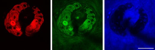
A new kind of biomedical imaging developed at Harvard is allowing researchers to capture video at scales never before seen, allowing for streaming footage at the subcellular level. The new technique, based on stimulated Raman scattering (SRS), can capture video of red blood cells squeezing through capillaries.
The technique, with its unprecedented speed and precision, will make for a nice complement to MRI, which captures fantastically detailed static images of organs and other tissues deep within the body but isn’t great for capturing unfolding events inside the body. SRS microscopy allows for the video imaging of proteins, lipids, and water within cells without the need for chemical labeling or other intrusive methods of tagging certain subcellular objects. Put another way, we’ve never been able to capture video this at this scale this well.
The Harvard team has already used SRS microscopy to study the migration of topical medications through the skin, and it could shortly begin aiding in surgeries to remove tumors in which samples have to be removed and the histologically examined – a process that takes 20 minutes or so while the patient lays on the operating table. SRS microscopy could do the same analysis in real time in the operating theater. Coupled with endoscopy, it could also create 3-D sections of tissues layer-by-layer, giving clinicians a better view of what’s going on.
Check the video below to see just how up-close the new video technique is. It shows real time blood flow through a Y-shaped junction of capillaries in a mouse ear, though we’re fairly certain no animals were harmed during the making of this film.