Before photography was developed, doctors could only depend on each other’s written descriptions and drawings to communicate what to look for in a scarlet fever rash, or how consumption affects the lungs. So a number of multi-talented physicians from the medieval and early modern periods published illustrated books that showed diseased people and body parts. (Yum.) We’ve collected a few of these drawings here. Consider them a companion to our seven favorite vintage anatomy drawings, which show healthy skeletal, vascular and other body systems.
These and other disease drawings are also on display until December 20, 2013, at the Indiana University Bloomington’s Lilly Library.
Click here to enter the gallery
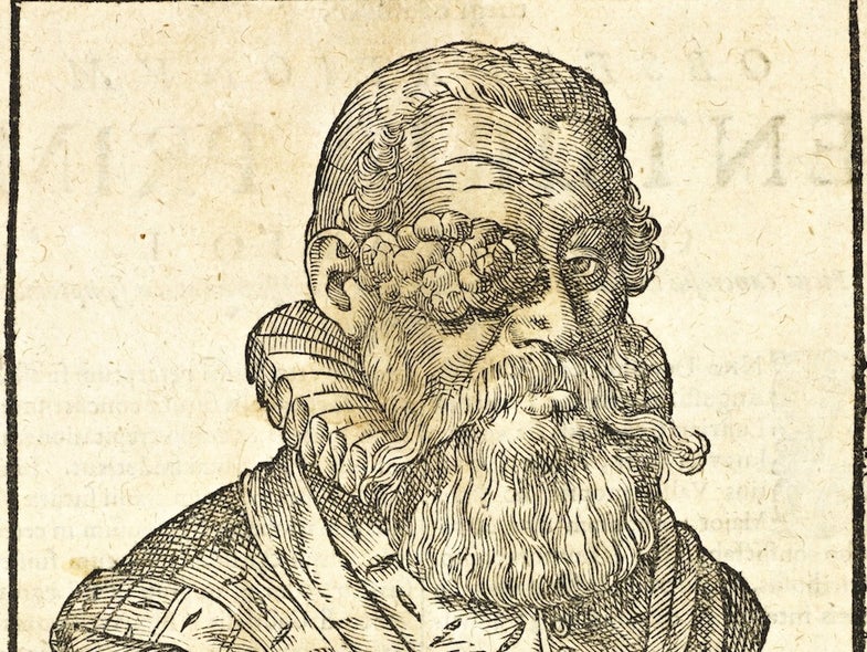
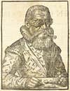
Ruling Through Adversity
Guilhelmus Fabricius Hildanus was known as the father of German surgery, performing procedures “that would be classified today as neurosurgery,” according to a paper about him published last year in the journal Child’s Nervous System. In this woodcut, Hildanus portrayed Claude Mayor, the ruler of a small Swiss town called Lutry. Mayor had an eye tumor.
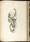
Osteographia Baby
This is an image from English surgeon William Cheselden’s Osteographia, or the Anatomy of the Bones. We haven’t been able to track down what, exactly, it depicts, although something seems wrong, doesn’t it? Cheselden put a lot of money into Osteographia, which was large and featured dozens of engravings, but it did not sell well. Costly and time-consuming to make, anatomical atlases of the time were often financial failures, the U.S. National Library of Medicine notes. If only Cheselden could have known—copies of his book now sell for tens of thousands of dollars.
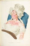
Miss Scarlet In The Bedroom With A Bacterium
This illustration of a scarlet fever rash, made by a French dermatologist, also happens to show some fashions of the time.
This Is Your Lung On Tuberculosis
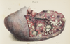
Carswell was a pathologist in London. Here he illustrates a lung from a patient with pulmonary tuberculosis, which was prevalent in industrialized countries in the 19th and early 20th centuries. A scientist first identified the microbe that caused the illness in 1882, long after Carswell made this drawing.
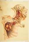
‘Hodgkin’s Malady’
This is an early illustration, made by two English pathologists, of the cancer now called Hodgkin’s lymphoma. The cancer causes cells in the lymph nodes to grow abnormally and impairs the immune system.